Sponsored by HORIBAOct 18 2013
Raman Microscopy has proven to be an extremely useful technique in the analysis of solid phase particles and surfaces. With the latest automated technology, analysts are able to explore surface structure and contaminants easily. Law enforcement agencies and forensic scientists need to verify the presence of explosives on a surface and characterize the distribution of individual elements in an explosive material.
Earlier, limited information have been acquired using destructive analytical techniques such as mass spectroscopy and chromatography and qualitative methods such as AFM. Nevertheless, the requirement for destructive and extensive extraction procedures is one of the shortcomings of these methods. In addition, little or no direct characterization of the sample is only possible with them.
Alternatively, the Raman microscope is capable of providing non-destructive, in-situ, highly specific spectral and distribution information. This article discusses the application of a LabRAM 1B confocal Raman microscope from HORIBA Jobin Yvon to analyze a surface containing an explosive composed of two typical components, PETN and RDX.
Instrumentation
The LabRAM 1B confocal Raman microscope is a robust single stage Raman spectrometer system equipped with integral microscope, holographic notch filter technology, and CCD camera. The compact system is integrated with the patented Laser line scanning facility and an automated motorized XY scanning stage to acquire a full 2D Raman map. It is also coupled with an internal HeNe laser and a secondary 532nm laser, although only the HeNe laser is used for this work.
With unprecedented optical performances in real confocality and sensitivity, the LabRAM can construct a Raman mapped image of the explosive material with a spatial resolution of roughly 1µm. The LabRAM 1B confocal Raman microscope with integral confocal microscope, low and high resolution dual grating option, internal HeNe laser, CCD video camera and automated motorized XY scanning stage is show in Figure 1.
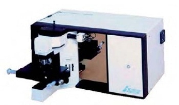
Figure 1. The Confocal LabRAM 1B Analytical Raman Microscope System from HORIBA Jobin Yvon.
Experimental Procedure
The LabRAM system was used to acquire digitized white light video images to visually analyze the sample surface for determining the presence of possible explosive contamination. By setting up 2-D Raman maps, a full analysis of the defined area was produced and complete Raman spectra were acquired from each micron of the surface. Spectral characteristics of the different explosive components were determined and a Raman image characterizing the localization of the active explosive components and the binder was then subsequently produced by the Windows LabSpec software.
Experimental Results
The digitized white light video image of the sample area analyzed is illustrated in Figure 2, apparently showing the presence of some contamination on the surface. The box represents the defined area for the 2-D Raman map, with a size of 20 x 30µm and a step size interval of 0.5µm. A Raman image was generated using the v(C-H) stretch Raman bands in the spectral region 2800-3200cm-1, showing the localization of all components of the suspect material.
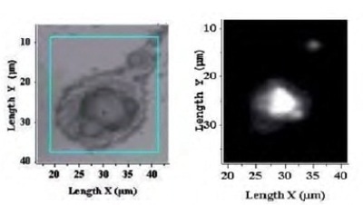
Figure 2. White light video image (left) contaminated surface.
The typical spectra for each explosive component acquired are depicted in Figure 3. The localization of the different particles of each explosive component is illustrated by individually mapping and superimposing the components (Figure 4). The distribution of the inert material was then confirmed by mapping the binder (Figure 4).
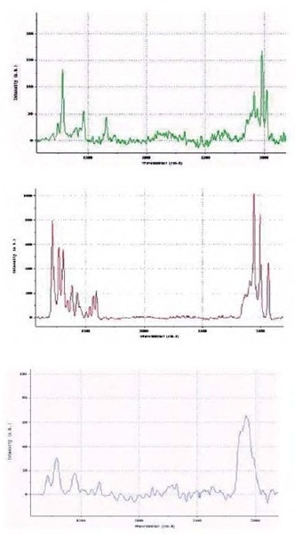
Figure 3. Raman spectra for the different components in the explosive material.
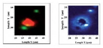
Figure 4. (left) Raman mapped image for the two active components, RDX (red), and PETN (green), and Raman mapped image for the inert binder (right).
Conclusion
The confocal Raman mapping results clearly distinguish the presence of three main components in the explosive material and confirm that they are RDX, PETN and binder. In addition, the results show a close positional correlation between the images produced by Raman mapping and the white light image.

This information has been sourced, reviewed and adapted from materials provided by HORIBA.
For more information on this source, please visit HORIBA.