Sponsored by HORIBAOct 28 2013
Simultaneous probing of an object using an atomic force microscope (AFM) and a vibrational technique like Raman Confocal spectroscopy to study surface topography and composition, respectively, is gaining interest in a range of applications, from biomaterials and nanomaterials to in-vivo biological cell study. Acquiring the vibrational signature with a spatial resolution of around 100nm provides key insights into the exotic properties of nanomaterials owing to possible confinement effects.
The correlation between the highly resolved Raman signal and the topographical characteristics enables localizing defect areas of interest that can be induced either mechanically (using an AFM tip) or optically (using the confocal microscope). Exceeding the resolution limit of traditional optical microscopy by a combination of traditional Raman confocal microscopy and near field techniques is a difficult task that provides many benefits in terms of acquisition time resolution and spatial resolution.
Instrumentation
Veeco Instruments and HORIBA in partnership with the University of Bordeaux spectroscopy group developed a versatile platform (Figure 1) by combining a sophisticated Confocal Raman microscope and AFM in a transmission geometry. The two instruments can be controlled in a step-scan AFM/Raman acquisition mode, thanks to a sturdy mechanical coupling between them as well as communication protocol (Veeco Open Architecture Labview routine (Figure 2) and HORIBA protocol).
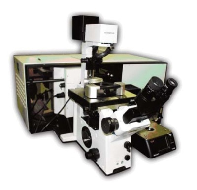
Figure 1. Combination of the Veeco Bioscope II with the HORIBA LabRAM HR spectrometer
Once in feedback, Raman acquisition is started by positioning the tip at a given XYZ position. In each XYZ (or XY Error, XY phase,…), position respective to a Raman Spectrum is acquired over a broad spectral range. The spacing and the acquisition times between the XY points can be controlled by users and rely on the spectral range probed. After repeating the above said procedure on a broad array of user-defined points, the sample is then moved in the XY position at the same time of aligning the focal point and tip. The feedback of the tip allows movement along the Z direction only.
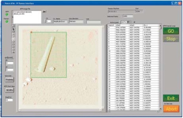
Figure 2. Veeco Open Architecture Labview routine: the initial AFM topography image is divided into discretized points.
Nanomaterial Characterization Using the Combined Setup
Single semiconductor nanowires with different composition, sizes and surface modifications are analyzed using the combined setup. The deposition of the samples takes place on the surface of a transparent substrate. For nanowires, the object can be mechanically stabilized at the surface by atomic layer deposition of aluminium oxide. AFM and Raman images of silicon nanowires deposited on a glass coverslip can be recorded simultaneously. The Raman spectra reveal local broadening and spectral shift, which can be correlated with the material properties. Scheme of the Bioscope II/LabRAM setup combination and the spectra of silicon wires are shown in Figure 3.
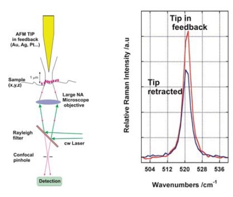
Figure 3. Scheme of the Bioscope II/LabRAM setup combination and Spectra of Silicon wires
Superimposition of the software applied for controlling the AFM and the Raman confocal microscope is shown in Figure 4. Both software interfaced by the Labview routine through TCP-IP protocol.
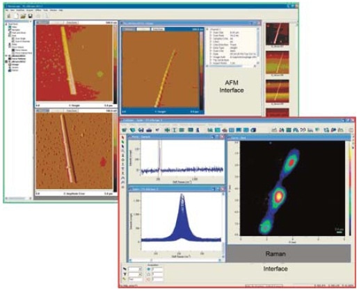
Figure 4. Superimposition of the software applied for controlling the AFM and the Raman confocal microscope
Tip Enhanced Raman Spectroscopy
Tip enhanced spectroscopy involves interaction of a metallic coated tip with the object being analyzed. The interaction between a focalized laser with the proper polarization and the metallic tip improves the EM field by the excitation of the tip’s plasmon band. Since the metallic tip is within the near field of the sample, the excitation with the proper wavelength can improve the field significantly, resulting in larger Raman signal from the sample.
This also improves the spatial resolution of the optical measurement because the enhancement is originating from the small tip apex. If the tip is positioned far away from the sample, the microscope objective is able to acquire only the far field signal. The variation between the two Raman spectra with the AFM tip in near field or far away from the sample provides the near field contribution, and consequently the tip enhanced contribution. Superimposition of the softwares
Conclusion
The combination of the HORIBA LabRAM HR and the Veeco Bioscope II is flexible enough to be used either independently or simultaneously. This sophisticated platform enables the analysis of both chemical and morphological data at the same sample location as well as the analysis of nanostructures with an xxx spatial resolution. The following are the key advantages of LabRAM HR:
- High resolution Raman spectrometer, with wide selection of gratings and lasers excitations between UV and NIR
- Option for Dual CCD detection and monochannel (InGoRs, PMT...) detection
- True confocal microscopy with software adjustment of the confocal aperture
- A broad range of X47 mapping capabilities and options
The following are the key advantages of Bioscope II:
- Near IR Laser diode
- Mechanical stability
- Independent Z and XY scanner for a higher frequency response

This information has been sourced, reviewed and adapted from materials provided by HORIBA.
For more information on this source, please visit HORIBA.