Branching is a critical structural parameter of many natural and synthetic polymers and considerably impacts their properties. These properties comprise viscosity and rheological behavior of polymer solutions and melts, thermodynamic properties, and various mechanical properties.
Quantitative data about branching topology is very important to gain a better understanding of polymerization processes and also to develop innovative polymer-based materials with improved properties.
Using methods like asymmetric flow field flow fractionation (A4F) or size exclusion chromatography (SEC) to characterize branching is not possible without a multi-angle light scattering (MALS) detector. To this end, branching information can be explained by means of actual experimental results obtained with Wyatt Technology instruments. This article describes the most common methods employed for the characterization of branching.
Branching Characterization
The history of branching characterization started with a prominent article by Zimm and Stockmayer who introduced a parameter called the branching ratio, g.
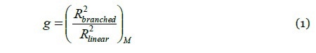
Where R2 denotes the mean square radius of branched and linear macromolecules that exhibit the same molar mass (M). g is directly related to the number of arms in star-branched polymers or to the number of branch units in arbitrarily branched polymers. g is ≤ 1, where the equality sign is applicable for linear polymers. When the value of g is lower, the degree of branching will be higher. For instance, a value of g ≈ 0.1 denotes a highly branched structure.
After a decade of the definition of g, Zimm and Kilb introduced another branching ratio based on the intrinsic viscosity.

where [η] indicates the intrinsic viscosity of linear and branched polymer molecules which exhibit the same molar mass. The relationship between g and g’ is explained through the ‘draining parameter’ e:
g' = ge (3)
The parameter e is anticipated to differ in the range of 0.5 - 1.5, while a value of e ≈ 0.7 may be utilized to recalculate g and g’.
From equations 1 and 2, it is evident that in order to characterize branching, information is needed about the molecular size and molar mass. Here, the MALS detector proves useful as it offers both sets of information concurrently and independently. The detector is normally joined to an analytical separation method - commonly SEC - to ascertain branching parameters as a virtue of molar mass. The parameters of interest are the number of branch units per molecule and the branching ratio.
SEC elution volume and intrinsic viscosity are the two most effective alternatives. The former can provide g using the approach of Yu and Rollings, while the latter can be utilized to calculate g’ using Equation 2.
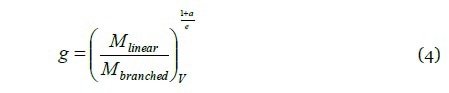
where ‘M’ stands for the molar mass of branched and linear molecules eluting at the same elution volume V, and ‘a’ represents the exponent of Mark-Houwink equation for linear polymers.
Techniques for Characterizing Branching
The results presented here were obtained using several instruments from Wyatt Technology: the DAWN HELEOS® MALS photometer; the Eclipse A4F system; a refractive index detector Optilab T-rEX® refractive index detector; and the ViscoStar® online differential viscometer. The data were processed with ASTRA® software. Also from Wyatt, the Tetrahydrofuran was utilized as a solvent for A4F and SEC methods.
A number of methods can be used to obtain branching ratio-versus-molar mass plots and they include radius method, mass method, and viscosity method. The radius method calculates g from the conformation plot using Equation 1; the mass method determines g from the molar mass–versus–elution volume plot using Equation 4; and the viscosity method calculates g’ from the Mark-Houwink plot using Equation 2.
The radius method is the most accurate method to characterize branching; however, it is restricted to polymer molecules with RMS radius < ≈ 10nm. In contrast, the mass method is ideal for small polymers for which the RMS radius cannot be determined. One major advantage of the viscosity method is that it provides high sensitivity with respect to branching and also offers the possibility to measure over a wide range of molar masses down to 1000g/mol.
Experimental Results
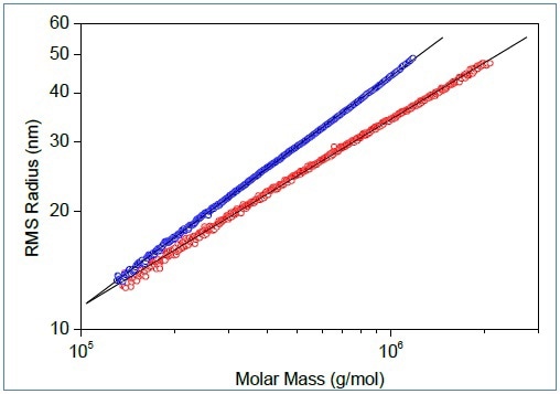
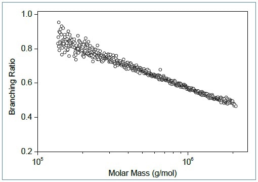
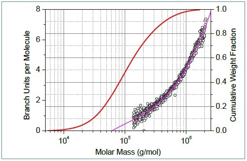
Figure 1. Conformation plots of linear (blue) and branched (red) polystyrene. Center: The corresponding plot of branching ratio versus molar mass. Bottom: The number of branch units per molecule plotted versus molar mass.
Figure 1 shows an example of the conformation plots of branched and linear polymers. One can easily identify branching from the slope of the plot. The conformation data help in determining branching. It is useful to overlay the plot of branching parameter with the collective distribution of molar mass. In Figure 1, it can be seen that ≈ 28% of molecules with molar masses less than ≈ 60,000g/mol do not have branched units. Remarkably, SEC-MALS is able to detect just a single branch unit in polymer chains.
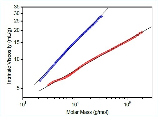
Figure 2. Mark-Houwink plots of linear (blue) and branched (red) poly(lactic acid). Bottom: Molar mass–versus–elution volume plots, same colors.
Figure 2 shows the characterization of branching for tiny polymer molecules, comparing molar mass-versus-elution volume and Mark-Houwink plots of branched and linear polyesters based on lactic acid. This polymer is bio-compatible and bio-degradable and can be used as a drug delivery material; its capability to swell and release an active compound can be managed by the degree of branching.
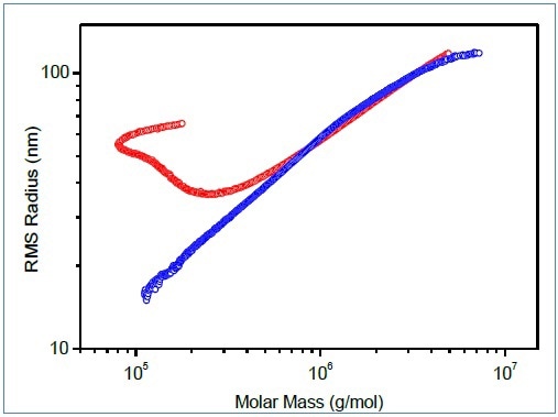
Figure 3. Conformation plots of polymer containing branched macromolecules determined by SEC-MALS (red) and A4F-MALS (blue).
For polymers demonstrating irregular conformation plots, A4F has proved to be an excellent separation technique and offers superior conformation plots. Figure 3 shows the comparison of conformation plots acquired by A4F-MALS and SEC-MALS.
Conclusion
Wyatt MALS photometers not only help in determining branching ratio g, but also help in calculating the number of branch units per molecule. Using an online viscometer, SEC-MALS set-up can be completed to ascertain Mark-Houwink plot. Moreover, by means of Mark-Houwink plot one can characterize smaller branched polymers. For separation of branched polymers, A4F method provides excellent separation and produces accurate conformation plots and values of g.
References
- Reprinted with permission from "Branching revealed: Characterizing Molecular Structure in Synthetic and Natural Polymers by Multi-Angle Light Scattering." White paper by Wyatt Technology.
- Polymer Characterization Webinar: Branching revealed: Characterizing Molecular Structure in Synthetic and Natural Polymers by Multi-Angle Light Scattering. Speaker: Dr. Stepan Podzimek.

This information has been sourced, reviewed and adapted from materials provided by Wyatt Technology.
For more information on this source, please visit Wyatt Technology.