Scientific advancements are often inspired by nature. For instance, penicillin was discovered in mould, aspirin was made from willow bark and solar cells imitate the photosynthetic process. Now, vertebrates and invertebrates are being studied in detail to develop innovative medicinal compounds. Unlike mammals, marine algae and phytoplankton synthesise different types of omega-3 (ω-3) polyunsaturated fatty acids (PUFAs).
PUFAs are natural products that regulate important metabolic mechanisms such as heart and brain activity. They are known to have anti-cancer activity and to reduce inflammation. A diet that includes fish is the main source of PUFAs for human beings. This is because fish themselves feed on microorganisms rich in ω-3.
The term ‘ω-3’ describes how these molecules contain a double bond on the third carbon from the chain terminus. The ω-3 class includes a wide range of molecules which show a lot of variation in terms of the extent of saturation and the chain length. α-linolenic acid (ALA), eicosapentaenoic acid (EPA) and docosahexaenoic (DHA) are the three most important ω-3 molecules found in nature (Figure 1).
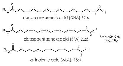
Figure 1. Structures of DHA, EPA and ALA molecules
EPA serves as a precursor to several eicosanoids; a family of important biological signaling molecules. Eicosanoids are involved in critical feedback loops in blood regulation such as the activation of white blood cells and the aggregation of platelets.
A key part of the skin, cerebral cortex and retina; DHA is an essential fatty acid that can be directly metabolized or produced in vivo from ALA. It regulates brain function, enhances cardiovascular health, optimizes the efficiency of chemotherapy, slows the progression of Alzheimer’s disease and prevents the growth of some cancers.
Today, a range of PUFA supplements are available on the market as human diets do not contain adequate amounts of ω-3 molcules. This article uses benchtop 60MHz and standard 400MHz 1H NMR spectroscopy to highlight the variations between four supplements; which include wild salmon oil, shark liver oil, cod liver oil, and ω-3 supplements.
Omega-3 Supplements
A standard ω-3 supplement will include PUFA, fish oil concentrate (PUFA-EE), EPA, and DHA. Fish oil concentrate is a synthetic product, and is exclusive to ω-3 supplements. It is produced via a transesterification reaction between ethanol (EtOH) and a PUFA triglyceride to give the ω-3s as ethyl ester derivatives. The reaction results in the elimination of the triglyceride chain as glycerol (Figure 2).
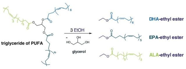
Figure 2. Transesterification reaction of a triglyceride of PUFA and ethanol.
This process makes it easier to purify, refine, and concentrate the PUFAs. However, the derivatives obtained are less bioavailable because they have to be changed again to the triglyceride form in vivo. The ethyl ester derivitive's spectra at 400 MHz and 60 MHz 1H NMR show a quartet at δ 4.09ppm (-CH2-0C(=0)-). A triglyceride linkage is not observed at δ 4.21ppm (Figure 3).

Figure 3. Ethyl ester derivative giving rise, in 400 MHz and 60MHz 1H NMR, to a quartet at δ 4.09ppm (-CH2-0C(=0)-) with a prominent absence of the triglyceride linkage at δ 4.21ppm.
| % |
60 MHz |
400 MHz |
%Difference |
Olefin
-CH=CH- |
22.87 |
22.84 |
0.12 |
ethyl ether
-CH2O- |
5.18 |
5.13 |
1.09 |
bis-allylic
=CH-CH2-CH= |
18.54 |
18.46 |
0.44 |
α and β
-CH2- |
21.47 |
21.37 |
0.46 |
alkyl
-CH2- |
24.54 |
24.14 |
1.69 |
Terminal methyl
-CH3 |
7.39 |
8.06 |
-8.31 |
| Total % |
100.00 |
100.00 |
n/a |
Wild Salmon Oil
Wild salmon oil is a less processed supplement. The label in a wild salmon oil will usually include a list of DHA, EPA and wild Alaskan salmon oil. High amounts of saturated fats are associated with an increased risk of cardiovascular disease. Evidence indicates that monounsaturated fatty acids regulate the normal growth of cells and help in reducing inflammation.
Western diets often contain more ω-6 and less ω-3, however, a high ω-6/ω-3 ratio can negatively impact human health. Triglyceride is a natural form of fatty acids and it is present in salmon oil. Since salmon oil is not as processed as other supplements it has a higher quantity of monounsaturated fatty acids such as oleic acid and saturated fatty acids like palmitic acid. It also contains the required amount of PUFAs such as linoleic acid (DHA).
Figure 4 shows the structure of DHA, palmitic, linoleic and oleic acids, and Figure 5 shows the 60 Mhz and 400MHz 1D 1H NMR spectra for wild salmon oil.
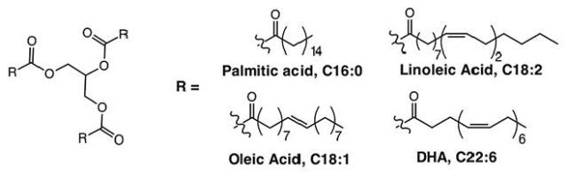
Figure 4. Structures of DHA, palmitic, linoleic and oleic acids.
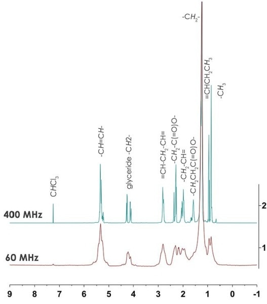
Figure 5. 400 and 60MHz 1D 1H NMR spectrum for wild salmon oil with % difference.
| % |
60 MHz |
400 MHz |
%Difference |
Olefin
-CH=CH- |
12.25 |
|
1.47 |
glyceride
-CH2- |
3.91 |
3.79 |
3.18 |
bis-allylic
=CH-CH2-CH= |
7.10 |
7.00 |
1.44 |
α and β
-CH2- |
20.30 |
20.50 |
-1.02 |
alkyl
-CH2- |
47.02 |
47JMÊ |
-0.94 |
Terminal methyl
-CH3 |
9.41 |
9.16 |
2.82 |
| Total % |
100.00 |
100.00 |
n/a |
A good agreement is seen between the 400 MHz and 60 MHz data acquired for the percent composition of the nutritional supplement, and this data is similar to that obtained for the ω -3 supplements. While 400 MHz NMR can be used to establish the ω-3: ω -6 acid ratio, inadequate resolution on the 60 MHz spectra makes it difficult to accurately measure the ratio.
Despite this fact, both saturated and monounsaturated fatty acids can be viewed through 1) a higher number of alkyl -CH2- components (~47% cf., 24% in ω-3 supplement) and 2) a higher olefin to bis-allylic ratio (1.7 cf., 1.2 in to ω -3 supplement). The multiplet centered at δ 4.21 ppm makes it possible to view the triglyceride linkage.
Cod Liver Oil
A standard cod liver oil supplement will contain vitamin A (palmitate), vitamin D (cholecalciferol) and cod liver oil. Cod liver oils are a good source of ω-3, predominantly DHA and EPA, and are similar to omega-3 and wild salmon oil supplements. These are free fatty acids, and occur naturally in their triglyceride form. Also, their composition is analogous to that of salmon oil which includes large quantities of saturated fatty acids (such as stearic and palmitic acids) ω-6's like linoleic acid, and monounsaturated ω-9's like oleic acid.
Significant amount of vitamins A and D3 are present in cod liver oil. However, while vitamin A is present in a high concentration resonances cannot be seen in either spectrum. Vitamin D is found at a lower concentration which is below the detection limit in both the 400 MHz and 60 MHz 1D 1H NMR spectrum. These components can be detected through a more sensitive method. Figure 6 shows the 400 and 60MHz 1D 1H NMR spectrum for cod liver oil.
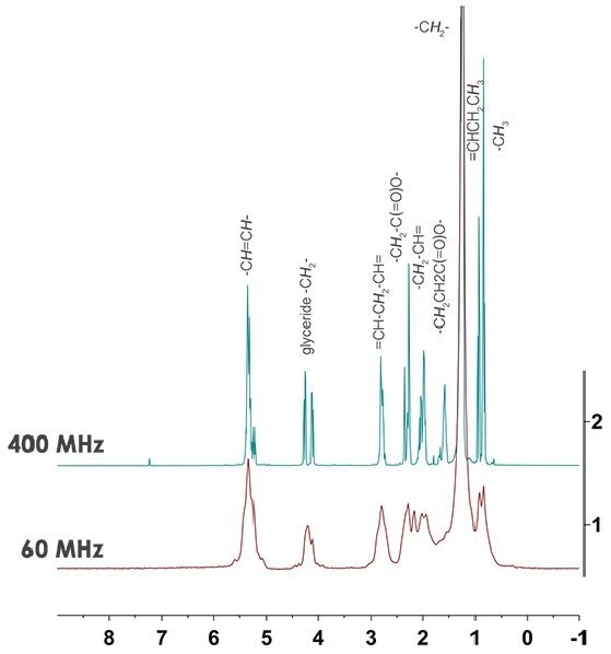
Figure 6. 400 and 60MHz 1D 1H NMR spectrum for cod liver oil with % difference.
| % |
60 MHz |
400 MHz |
%Difference |
Olefin
-CH=CH- |
ll.46 |
12.08 |
-2.60 |
glyceride
-CH2- |
4.00 |
4.00 |
-0.12 |
bis-allylic
=CH-CH2-CH= |
6.80 |
6.84 |
-0.50 |
α and β
-CH2- |
20.53 |
20.90 |
-1.75 |
alkyl
-CH2- |
48.40 |
47.60 |
1.67 |
Terminal methyl
-CH3- |
8.81 |
8.89 |
-0.95 |
| Total % |
100.00 |
100.00 |
n/a |
The presence of retinyl palmitate, i.e. vitamin A, in supplements can present a major issue because it is not well tolerated by humans. Although essential for many basic functions, vitamin A is a fat-soluble vitamin and is likely to accumulate in fat tissues.
People should take cod liver oil only under the supervision of a doctor, and must limit and regulate other vitamin A sources in their diets. Toxins are generally removed by the liver, and trace amounts of contaminants such as MeHg, PCBs, PBDEs, and PCDDs may be present (Figure 7). If these contaminants exist in the samples their low concentration would make it difficult to detect on a 400 MHz or 60 MHz spectrum.
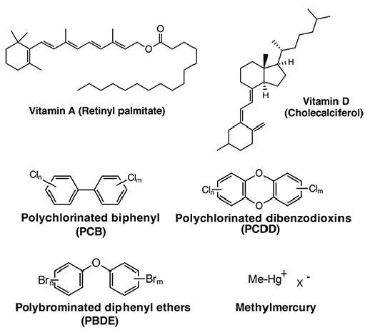
Figure 7. Structures of vitamins A and D, PCB, PCDD, PBDE and MeHg.
Shark Liver Oil
This oil is obtained from specific species of deep sea sharks. It does not contain much ω-3 PUFAs, however it is still erroneously classified as a PUFA supplement. The liver of a shark has a special composition because it serves as a swim bladder. It contains oils that boost the fish’s buoyancy and these oils contain mostly squalene.
Squalene, a terpenoid, is used as a precursor for in vivo steroid development. Shark liver is the chief source of squalene, though other natural sources are also available, namely amaranth and vegetable oil. Squalene is known to reduce cancer risk, shrink existing tumors, and also have anti-carcinogenic activity. Whilst squalene has a different bioactivity, it provides similar health benefits to PUFAs.
However, shark liver oils contain pristane and toxins such as PCBs and PBDEs. There is no proof of aromatic protons from PBDE or PCB contaminants in the 400 MHz or 60 MHz spectrum, and this indicates that these contaminants are at a level lower than the detection limit or not present altogether. However, alkyl impurities were detected in the allayed samples.
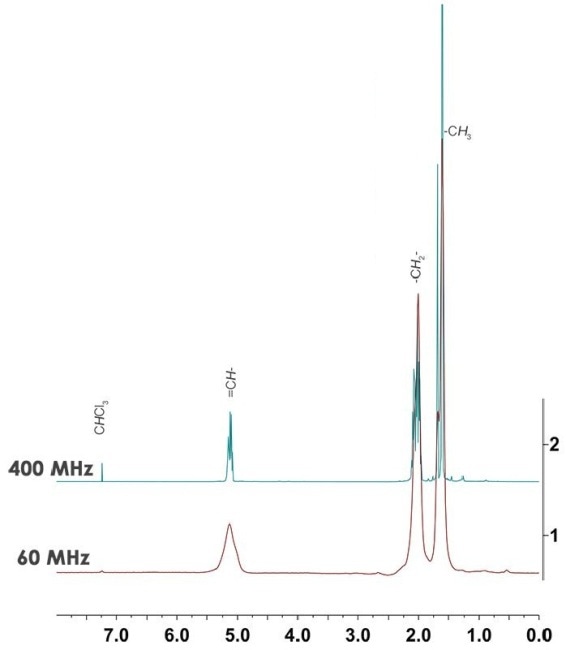
Figure 8. Spectra exhibit evidence of alkyl impurities at both high and low fields in the 5.0-1.4 ppm range.
In Figure 8, the spectra show the presence of alkyl impurities at low and high fields in the 5.0 - 1.4 ppm range. This can be attributed to the remaining pristine, a saturated terpenoid substance that is the second major source of oil in shark liver.
Pristine can cause rheumatoid arthritis in humans and, plasmacytosis and auto-immune disease in mice. A more sensitive method such as high-performance liquid chromatography (HPLC) or mass spectrometery should be used to establish whether this impurity is a residual solvent or pristine.
Conclusion
From the results, it is evident that the constituents of fish oil supplements can be effectively analyzed using the standard 400MHz and benchtop 60MHz 1H NMR spectroscopy.
This information has been sourced, reviewed and adapted from materials provided by Nanalysis Corp.
For more information on this source, please visit Nanalysis Corp.