Today’s advanced digital microscopes feature sophisticated digital imaging technology and superior quality optics, enabling even novice users to instantly capture high-quality images and produce reliable results.
These instruments offer a range of observation methods, intuitive magnifying operation, easy observation, and reproducibility. Operating these microscopes is as easy as using a tablet or smartphone.
Advanced digital microscopes offer effective solutions to a number of common microscope problems faced by users of traditional optical and digital microscopes. This article discusses 10 traditional microscopy challenges and the corresponding solutions that can be achieved with today’s digital microscope technology.
Issue 1: Specific Details Need to be Seen on Challenging Samples
It is not possible to observe low contrast samples in most imaging cases. Sample surface conditions cannot be clearly observed with low contrast and low reflective difference even after magnification. In other cases the sample surface can be disturbed by glare caused by halation, so performing a clear observation is a challenging prospect. Carefully adjusting illumination can eliminate this glare, but it is a time-intensive process and may still not yield satisfactory results.
Solution: Optimized Digital Imaging Processing
Today’s digital microscopes use sophisticated digital image processing that help to clearly observing surface conditions, which are otherwise difficult to observe with traditional optical microscopes (Figure 1). For instance, the Evident DSX series digital microscope comes with the High Dynamic Range (HDR) function that enables optimal observation even with a low contrast image, by integrating several images captured at different exposures (Figure 2).
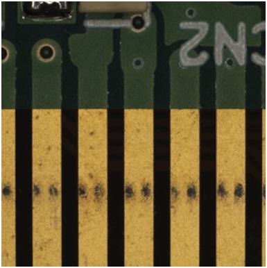
Figure 1. When performing inspection with a traditional microscope, it is often impossible to image the inner pattern of the circuit board.
Samples that may require multiple pieces of equipment earlier for accurate observation can now be clearly investigated with a single system, thanks to the HDR function. The DSX also features a proprietary image processing system called WiDER to address high-contrast problems with a single click, thus eliminating glare. WiDER works efficiently with live images as it does not require laborious adjustments to illumination.
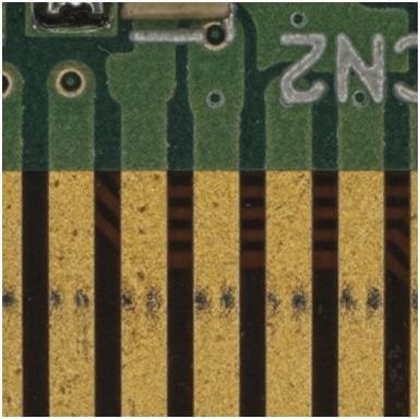
Figure 2. Using a digital microscope, a live image can be displayed using HDR observation to combine multiple images with different exposure times. (Image created using the Evident DSX510.)
Issue 2: A Large Sample Area Needs to be Observed in High Resolution
A sample cannot be viewed in its entirety at high resolution using traditional microscopes. Collecting an overall image of a sample becomes difficult as the observation field is narrowed due to an increase in the magnification.
Solution: Panoramic Imaging
Nothing comes “outside the field of view” in the latest digital microscope technology, thanks to panoramic imaging. With this technology, modern digital microscopes are capable of automatically stitching the images into a single seamless view through stage movement, thus achieving high resolution throughout all image regions even with larger samples. Panoramic imaging retains the original field while providing close-up clarity in extended focus, 2D, 3D or any combination of the three, compared to traditional microscopes that decrease field area with increasing magnification.
Issue 3: All Areas of an Uneven Surface Need to be in Focus at the Same Time
Traditional microscopes can sometimes only achieve clear focus on a portion of a sample with an uneven surface. The limitation of depth of focus makes it highly challenging to obtain clear focus on the whole sample, as the depth of focus becomes shallower with increasing magnification.
Solution: EFI (Extended Focal Image)
The DSX has an Extended Focal Image (EFI) capability that enables clear, in-focus images of entire samples to be obtained with a single click even if the sample surface is irregular. EFI simply moves the point of focus up or down in order to extract and integrate focused images. As a result, the focus is maintained across the entire surface area of the sample, enabling accurate analysis of uneven surfaces.
Issue 4: Sample Features Need to be Determined, Characterized, and Measured in 3D
The exact features of a dimensional sample can be difficult to determine based on a 2D image because of the plain view observation of an optical microscope. In certain cases, operators have to make their best estimate for height variations in samples or whether sample unevenness is convex or concave.
Solution: 3D Imaging
The ability of today’s digital microscopes to deliver 3D imaging of a sample with a single click provides an accurate view of the sample and enables the performance of an analysis from any angle (Figure 3). With accurate height data, in-depth 3D images enable viewing and measuring sample features or unevenness (Figure 4). It is also possible to measure height differences, so accurate sample analysis can be performed.
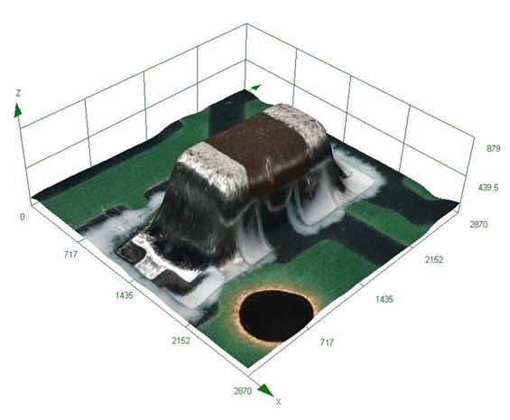
Figure 3. 3D image of surface capacitor. (Image created using the Evident DSX510.)
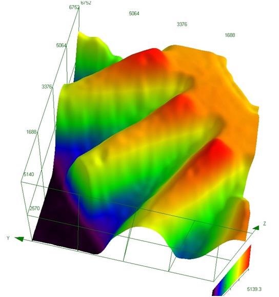
Figure 4. 3D image for height data of gear. (Image created using the Evident DSX510.)
Issue 5: Operators with Varying Skill Levels Need to Perform Similar Tasks
Conventional optical microscopes involve a series of complex operations and adjustments in order to switch between observation modes. Ascertaining the necessary image features for the task in hand requires the right level of expertise, otherwise it is a challenging process. Modifying the observation modes often needs special filers to be inserted, aperture stop and illumination to be adjusted, and much more.
Solution: One-Click Observation Modes
With the latest digital microscopes, operators of all levels can easily select the observation mode suitable for their applications with a simple click. All the user has to do is choose their optimal image from a set of acquired image options. The observation mode can be adjusted based on the selection. This suggests that both experienced and novice operators can perform the same types of high-level analysis.
Issue 6: Reproducible Measurements are Needed from Multiple Operators
Many settings and adjustments are required for traditional microscopes, so it can be a challenging process for multiple operators to perform an analysis under identical conditions. These conditions can start to change based on the operators and their specific method, causing major variations in images and position of microscope points. This can cause serious consequences in materials testing, research and development, or QA/QC.
Solution: Digital Repeatability
A fully digital microscope allows recording and referencing of all image acquisition and observation conditions at any time, including observation methods, stage coordinates, etc. This capability allows any operator to easily reproduce any analysis, ensuring inspection under the same settings and conditions. It is possible to recall image capture conditions with a single click. The system can further direct the user to carry out the same routine measurements via a guided interface.
Issue 7: Optical-Quality Imaging is Needed from a Digital Microscope
There is still a perception that an optical microscope is employed for high-quality images with higher resolution and natural color, while a digital microscope provides ease of operation. Most operators still classify their microscope applications between the two. This not only results in a waste of time, but also affects continuity of observation between optical and digital images.
Solution: Dedicated Lenses
Sophisticated digital microscopes use dedicated lenses, which are a combination of long working distances, high NA, evenness of light intensity, and well-controlled aberration. These lenses are then integrated with Full HD cameras and HDR digital processing, reducing glare, and allowing real color reproduction. Distortion and flare are eliminated, which were previously not possible with conventional digital microscopes.
Issue 8: Guaranteed Measurement Accuracy is Required from a Digital Microscope
Conventional digital microscopes generally do not offer clear accuracy guarantees. Operators do not have any way of determining the scope of error of the instrument they are using.
Solution: Telecentric Optics
Digital microscopes, such as the DSX, employ the same telecentric optics used in measuring devices, eliminating the variation in measurement results. The size of the observation target is not changed even if there is a change in the point of focus, ensuring no difference between operators, and enabling measurements to be performed under extremely stable conditions.
Issue 9: Varied Observation Techniques Require Different Lens Setups
Conventional digital microscopes consist of an array of lenses for different uses. This means operators need to switch between the lenses for different observations. It is a time-intensive process to replace conducting lenses between different types of observations and different types of samples. Several steps are needed to determine the optimum lens configuration for each application.
Solution: Variety of Observation Modes with One Lens
Modern digital microscopes feature an objective lens, which can be utilized for a range of observation methods. Therefore, optimal viewing conditions can be created without lens replacement, and operators can switch between observation modes with a single click (Figures 5 to 9).
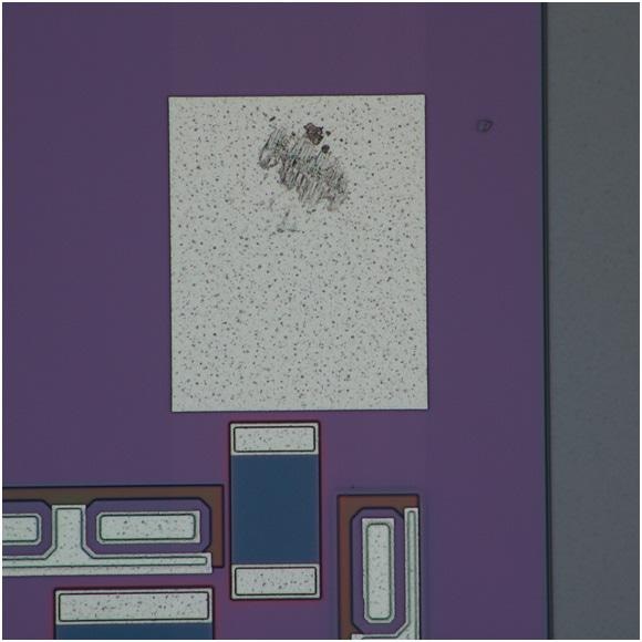
Figure 5. MEMS device image made with brightfield observation mode. (Image created using the Evident DSX510.)
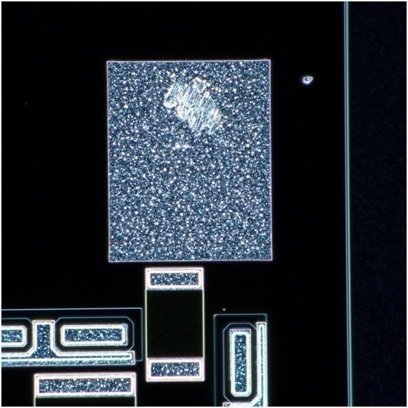
Figure 6. MEMS device image made with darkfield observation mode. (Image created using the Evident DSX510.)
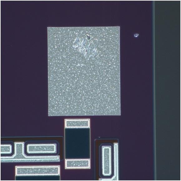
Figure 7. EMS device image made with MIX (brightfield plus darkfield) observation mode. (Image created using the Evident DSX510.)
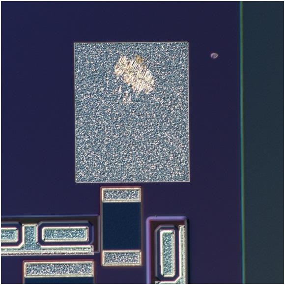
Figure 8. MEMS device image made with DIC observation mode. (Image created using the Evident DSX510.)
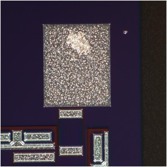
Figure 9. MEMS device image made with PO observation mode. (Image created using the Evident DSX510.)
Issue 10: Magnification Adjustments Require Manual Calibration
Manual calibration is required for many standard digital microscopes whenever there is a change in the magnification. When the incorrect calibrations are used to take images or measurements, the magnification indication and measurements will also become erroneous. As a result, it is necessary to repeat the whole process.
Solution: Automatic Magnification Recognition
Advanced digital microscopes, like the DSX, provide automatic magnification recognition using a motorized zoom system that reads the setting conditions of the mainframe in order to minimize human errors. The magnification setting is changed by altering the lens magnification automatically, eliminating the chance for measurement errors. The image area and current magnification information is updated when there is a change in the zoom magnification, further minimizing errors in the magnification indication and measurement values. In addition, the DSX system uses a dedicated standard to provide automatic calibration.
Conclusion
Today’s sophisticated digital microscopes deliver simple and intuitive operation, rapid and efficient observation, a range of image capturing techniques, and high-quality imaging and measurement results. They have options such as EFI and 3D imaging, programmed image capturing movie capturing, and panoramic image capturing, all integrated into an easy-to-operate interface.

This information has been sourced, reviewed and adapted from materials provided by Evident Corporation - Industrial Microscopy.
For more information on this source, please visit Evident Corporation - Industrial Microscopy