| Until recently, techniques for measuring birefringence have hardly altered with observations being made using standard polarising microscopes with white light and crossed polars, which can lead to coloured interference patterns in the sample image. To avoid this, the new Metripol technique uses a monochromatic light together with a rotating polariser and a circular analyser to carry out both qualitative and quantitative measurements of transparent microscopic specimens. it has useful applications in many areas, including the analysis of strain in industrial diamonds, phase transitions in crystals, and the analysis of collagen and hydroxyapatite distribution in bone. What is Birefringence? The phenomenon of birefringence (also known as double refraction) is a result of optical anisotropy, and can be described as the difference between two refractive indices for a given light beam, depending on the orientation of the polarisation of the incoming light. Birefiringence is displayed by a broad range of materials, including all crystals (except those of cubic symmetry), liquid crystals, glass and plastics subjected to mechanical strain. it also occurs in materials in which the underlying crystal structure may have its atoms distributed in such a way that it forms an anisotropic structure causing optical anisotropy. How Is Birefringence Observed in a Normal Polarising Microscope? The birefringence colours seen in a normal polarising microscope arise from a combination of three effects - intensity distribution of the light through the sample, the magnitude of the birefringence and the orientation of the indicatrix (φ). To get an exact image of the birefringence and determine its value, it is necessary to rotate the sample into a number of positions and determine the magnitude of the birefringence by inserting different compensating crystalline plates to effectively cancel the birefringence of the sample. For complex samples with numerous orientations this approach is very time consuming. The Metripol Technique Work carried out by Professor Mike Glazer at Oxford University, UK, into the relationship between crystal structure and physical properties, developed an entirely different approach to birefringence imaging. The research group wanted to create a system that could image, analyse and most importantly quantify birefringence. A collaboration with Oxford Cryosystems, UK, resulted in the Metripol emerging as a commercial analytical technique. How Does the Metripol Technique Work? The Metripol microscope produces quantitative birefringence data of samples in the form of images within a matter of seconds. The system incorporates a modified polarising microscope, rotating polariser, wideband adjustable wave plate and polariser (circular analyser). An integrated software suite is used to control the measurement process and for analysing the resulting images. Monochromatic light is used and images are collected using a CCD camera at different angles as the polariser rotates, which makes it possible to separate out the birefringence, orientation and transparency that are normally superimposed in conventional polarising microscopy. The software then generates three separate images, in which false colour is used to denote these components separately. The software also allows histograms and profiles through the images to be produced. Average values can be reported from selected regions of the images, so that the progress of a particular quantity at any place in the image can be studied - as a function of temperature, for example. The Multifile Facility A multifile facility allows the creation of a sequence of images that can be scanned through, in order to plot sequential values on a graph or to make an AVI video file of a process. Multifile creation can be automatically phased with an external source, such as a heating or cooling stage. Applications The Metripol system has already been used for a broad range of applications, from analysing collagen distribution in bone to studying optical properties of minerals, illustrating its versatility. Strain Analysis in Diamonds As a cubic crystal, diamond is not normally birefringent, and is optically isotropic. However, strain in diamond introduced by impurities, restrictions during growth or applied stress, makes the structure anisotropic and results in birefringence. Sources of Strain in Industrial Diamonds Producing diamond industrially using HPHT and CVD can cause the introduction of mechanical strain, which can significantly alter its physical properties causing twinning, crystal defects and weakness. However, in some situations the introduction of strain can actually strengthen the diamond, making it less susceptible to cleavage. Here, the Metripol technique has been used to generate both qualitative and quantitative information on different forms of diamond, including artificially grown diamond with nitrogen impurities, thin-film (CVD) diamond and diamond gemstone. Analysing Strain in Industrial Diamonds In the artificially-grown diamond shown in figure 1a, nitrogen has accumulated during the growth of the diamond. The diamond is grown as (111) plates (right, lower left, upper left) from a tiny seed (in the centre). At the sector interfaces there is a slight crystal-lattice misalignment, which allows for the incorporation of nitrogen. The misalignment, together with the presence of nitrogen, causes a build-up of strain leading to optical anisotropy. In the transmission image, the contrast in the centre is caused by absorption of light by the nitrogen. The birefringence image, figure 2b, indicates the magnitude of the strain in different parts of the diamond, with the purple colour representing the lowest values. The colours in the orientation image, figure 1c, indicate the orientation of one of the indicatrix axes and show that this axis points towards the centre of the diamond at all places. | 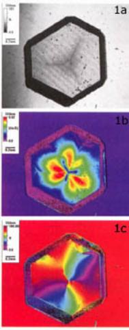
| | Figure 1. (a) Transmission image showing absorption at the centre caused by nitrogen. (b) Birefringence caused by strain associated with growth boundaries. (c) Orientation image, with strain orientations being marked by short lines in addition to the colour scale. | Phase Transition Studies The study of phase transitions is an important field of materials science, not only in its own right, but also for its importance in industrial applications. The Metripol was developed specifically for the purpose of following phase transitions, and as a result is perfectly suited to this application. Using the Metripol, crystallographic twins can be identified and, in some cases, the symmetry relating them can be determined together with the orientation of domain walls separating them. When a crystal is accurately cut and placed on a heating stage it is often possible to identify which phase the crystal is in, simply by determining the number of different twin domains visible in the crystal and the orientation of the indicatrix in each domain. Phase transitions can be accurately determined by the appearance of twin boundaries or from analysis of the change in birefringence as a function of varying temperature. Phase Transition in Sodium Bismuth Titanate The phase transition studies shown here have been carried out on Na0.5Bi0.5TiO3 (NBT) crystals, figure 2. Figure 2a illustrates the pure cubic phase 1 at 590°C showing low birefringence with random orientation. At 548°C, figure 2b, the tetragonal phase II begins to appear, especially in the orientation image. Figure 2c shows the pure tetragonal phase II ‑ notice the different birefringence in the central part of the cross feature. At 196°C, figure 2d, the rhombohedral-tetragonal coexistence region starts to form, and the rhombohedral phase III starts to appear in the orientation image. The end of rhombohedral-tetragonal coexistence occurs at 151°C, figure 2e. At this stage, the tetragonal phase II has nearly disappeared, and is only distinguishable in the orientation image. At 31°C, figure 2f, the pure rhombohedral phase III is visible, while residual signs of the tetragonal twin structure still remain in the orientation image. | 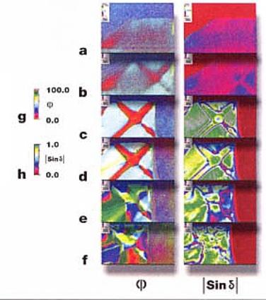
| | Figure 2. Phase transition studies using Metripol were carried out on Na0.5Bi0.5TiO3 (NBT) crystals. | Other Areas of Use for the Metripol System The Metripol system has been adopted by research groups in fields as diverse as diamond research, the study of polymorphic pharmaceuticals, engineering and biology. One of the most exciting developments has been in protein crystallography, in which research in collaboration with oxford and Warwick universities is starting to suggest that the Metripol may be a useful tool in determining crystal quality and early crystalline growth. Other exciting applications exist in earth sciences as an aid to identifying minerals in rock sections. Summary Qualitative and quantitative imaging of birefringence in transparent materials can add extremely valuable data to a broad range of studies in the fields of drug discovery, materials science and biological research. The Metripol imaging system offers an elegant solution to the problem of gathering such data. |