Sponsored by XradiaApr 22 2013
Carbon sequestration experiments have gained momentum as governments worldwide acknowledged them as an environmental initiative. Carbon sequestration shows promise to alleviate climate change by capturing carbon dioxide and preventing its emission into the atmosphere. Xradia 3D X-ray microscopes deliver high resolution imaging solutions, which enable a wide range of CO2 sequestration experiments.
Challenges
Storing carbon dioxide in geologic formations is one of the most potential techniques. This can be done in three ways: chemical reaction with rock minerals, dissolution in the fluids present in the rock pore space, or as immobile gas (due to capillary forces) within the pore space. Gaining insight into 3D microscopic fluid flow dynamics is essential to develop and optimize the interactions which ensure the carbon dioxide remains stored within the pore space of the rock.
Traditional techniques had its own limitation to directly investigate microstructure in 3D. Non-destructive methods like laboratory X-ray microcomputed tomography does not generally have the required resolution. Hence, scientists have often had to depend on computer simulations and artificial micromodels, or obtain pore-scale conclusions from macroscopic measurements.
Solutions from Xradia
Xradia is a leading provider of high-resolution X-ray imaging optics and laboratory-based 3D X-ray microscopes. The company has the world’s largest installed synchrotron base. The following are the laboratory-based 3D X-ray microscopes offered by Xradia:
- Submicron VersaXRM line that attains spatial resolution down to <0.7 µm
- Ultra-high resolution UltraXRM-L200 microscope that achieves spatial resolution down to 50 nm
The following key capabilities are offered by the combination of the VersaXRM family and UltraXRM to carbon sequestration studies:
- Resolution for pore network and clay porosity measurements
- Contrast for fluid imaging
- High resolution for in situ fluid flow analyses
The aforementioned capabilities of these synchrotron-quality laboratory systems enable carbon dioxide sequestration researchers to get in situ fluid flow observations and pore network morphology measurements to gain insight into relative permeability during drainage and imbibition.
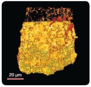
Figure 1. Pore network of chalky carbonate via UltraXRM-L200 (Sample courtesy of Robert Feidenhans'l, University of Copenhagen)
Resolution for Pore Network and Clay Porosity Measurements
Clay micro-porosity measurements and pore network geometry are crucial for developing predictive models for phenomena like gas trapping (Figure 1). Accurate flow modeling requires resolutions as high as submicron to tens of nanometers based on the rock formation and porosity. Xradia microscopes employ innovative two-stage magnification architecture to deliver high resolution with large field of view for submicron measurements of the clay microporosity and rock pore networks in comparatively large rock samples, as shown in Figure 2.
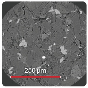
Figure 2. Resolution for pore network and clay porosity measurements
The VersaXRM and UltraXRM offer multiple imaging modes of many different field-of-view and resolution combinations. This capability is critical for gaining insight into heterogeneous rocks with a broad range of pore scales. It is possible to adequately characterize pores on all length scales through the use of multi-length scale imaging to derive a pore network model that is scalable up to a reservoir. The VersaXRM’s range of imaging modes provides Scout and Zoom imaging in heterogeneous rocks. It may be possible to identify and locate large pores in the higher throughput, lower resolution Scout mode. vuggy pore locations can be scanned by switching to a higher resolution objective in Zoom mode to image smaller pore throats that connect the pore to the remaining pore network.
Contrast for Fluid Imaging
Xradia platforms employ in-house detector systems that provide high contrast for low Z materials. The optimized contrast delivers three-phase imaging of air, unstained brine and sand, as shown in Figure 3. These image datasets can offer 3D measurements of relative fluid paths, particle contacts, and contact angles, which are critical data to understand carbon sequestration.
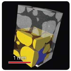
Figure 3. Contrast for Fluid Imaging Sand (yellow), Brine (blue), and Air (clear, brown) imaged in a 12.5 mm diameter aluminum tube
High Resolution for In Situ Fluid Flow Analyses
Limitations of conventional microCT overcome by Xradia 3D X-ray microscopes through their ability to provide high resolution at large working distances, enabling high resolution imaging of samples within in situ chambers and rigs, as shown in Figures 3 and 4.
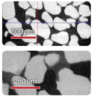
Figure 4. High resolution and contrast for in situ fluid experiments.
The VersaXRM-500’s large enclosure is specially designed to integrate labyrinths for accommodating cables and other feed-throughs in order to provide support for a variety of in situ chamber designs.
Conclusion
Hence, through the use of the high resolution 3D X-ray microscopes from Xradia, researchers can efficiently measure pore network morphology and observe in situ fluid flow in order to perform successful carbon dioxide sequestration experiments.

This information has been sourced, reviewed and adapted from materials provided by Xradia.
For more information on this source, please visit Xradia.