X-ray photoelectron spectroscopy (XPS), also referred to as electron spectroscopy for chemical analysis (ESCA), is an analytical technique used for characterizing the uppermost 10nm surface of materials. Employed in both academic and industrial research, the method characterizes surfaces with technological applications as varied as superhard, inorganic coatings or organic light emitting diodes.
The AXIS Supra+— Photoelectron Spectrometer
Kratos Analytical’s advanced AXIS Supra+ offers improved performance over its predecessor. The spectrometer can be used to acquire photoelectron spectra, giving quantitative chemical state information or high spatial resolution images, providing lateral distribution of elemental or chemical states at the surface. The AXIS Supra+ combines market-leading performance with ease of use.
With the help of the ESCApe integrated acquisition and processing software, the AXIS Supra+ performs to its highest capability and offers a user-friendly interface with the spectrometer. Moreover, the AXIS Supra+ is different from other spectrometers because instrument parameters and sample handling are fully automated via computer control. During analysis, a sample holder can be transferred and exchanged without user interference. This is realized through the coordination of the sample analysis chamber auto-stage and the Flexi-lock sample magazine.
Capabilities of the AXIS Supra+ : Large-Area, High-Sensitivity XPS
Kratos Analytical’s AXIS Supra+ has been improved for chemical state XPS. High transmission electron optics, together with efficient collection of photoelectrons ensures unmatched resolution and sensitivity at large analysis areas. In addition to the traditional scanned acquisition, spectra can be acquired in quick, unscanned snapshot mode within a second with the help of the 128 channel Delay-Line Detector (DLD).
Main attributes are given below:
- Data can be acquired quickly
- Light elements can be easily detected
- Includes scanned or snapshot spectral acquisition modes
- Exceptional signal-to-noise, even at reduced concentrations
High-Energy Resolution
The best possible energy resolution is the main requirement of any spectrometer. High-energy resolution is important to accurately measure the slight chemical shifts. Kratos Analytical’s AXIS Supra+ has a large 500-mm Rowland circle monochromated Al Kα X-ray source as well as improved electron optics, guaranteeing exceptional chemical resolution.
The high spectral energy resolution offers the following advantages:
- Assured energy resolution on conducting and insulating samples
- Explicit detection of chemical shifts
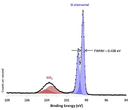
Si 2p region from native oxide on Si substrate acquired from large area with high energy resolution.
Small Spot, Selected Area Spectroscopy
Improved performance of the AXIS Supra+ small spot ensures high sensitivity in small spot mode. This is achieved by matchinng X-ray illumination with the chosen analysis area. An optional brite-X monochromatic source can also be specified for applications necessitating extremely high sensitivity performance from small selected regions.
Key attributes of the selected area spectroscopy are given below:
- Improved X-ray illumination for better performance of selected area spectroscopy
- Pre-defined small spot analysis regions, smallest analysis area 15 μm diameter
- Either the achromatic or monochromatic X-ray source can be used to acquire the selected area spectra
- Automated aperture and iris in the electrostatic lens column to create a virtual probe at the surface of the sample
- μ-boost acquisition mode for very high sensitivity at small area mode (option)
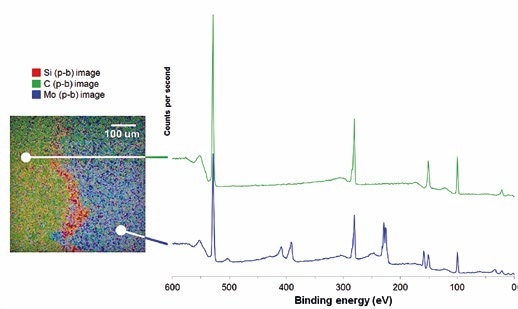
XPS image used to define a position for 27-μm selected area survey spectra.
Fast Parallel Imaging
XPS imaging is used to measure the lateral distribution of chemistry or elements at the surface. Rapid and high-spatial-resolution parallel images are acquired by the AXIS Supra+. The benefit of parallel image acquisition is that it is considerably faster and attains higher spatial resolution when compared to the more traditional rastered beam approach.
In addition, parallel imaging can be integrated with stage movements to obtain a “stitched” image; it can generate images over several millimeters with a spatial resolution of several microns.
Parallel imaging offers the following capabilities:
- Quantitative imaging—the exclusive spherical mirror analyzer as well as the delay-line detector can offer quantitative chemical-state images
- High-energy resolution, chemical-state imaging
- The ultimate spatial resolution of 1 μm at maximum magnification
- Spectromicroscopy—Spectra can be easily acquired from image datasets, thus offering a spectrum at each pixel
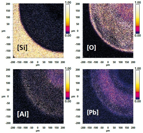
Quantitative images where the color scale represents a relative amount of each element present at the surface.
Multi-Technique Capability
Optional excitation sources comprise a high-energy Ag Lα monochromated X-ray source, enhanced helium discharge lamp for ultraviolet photoemission spectroscopy (UPS), and an achromatic Al/Mg X-ray source.
Moreover, the inclusion of a field-emission electron source adds scanning auger mapping (SAM), auger electron spectroscopy (AES), and secondary electron microscopy (SEM) ability to the instrument. These additional methods are coincident with the XPS analysis position, providing a complementary understanding of the sample. Notably, the inclusion of these methods does not affect the performance of the market-leading XPS.
Options like surface modification and sample preparation can be fitted on the introduction chamber, also called Flexi-lock. Such options include an air-sensitive sample transporter, sample heat and cool, crystal cleaver, broad spot ion source, and glove box.
It is possible to configure a third chamber to offer a dedicated UHV setting for surface science research. Usual configuration equips this third chamber with a manual stage as well as optional characterization methods such as quadrupole secondary ion mass spectrometry (SIMS), inverse photoemission spectroscopy (IPES), and low-energy electron diffraction (LEED).
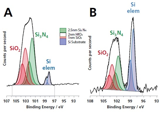
Si 2p spectra acquired from a thin-film multilayer sample (courtesy of IMEC) shown in the schematic figure using (A) monochromated Al Kα radiation and (B) monochromated Ag Lα. The greater information depth of the Ag Lα excited Si 2p spectrum is demonstrated by the larger Si elemental substrate component.
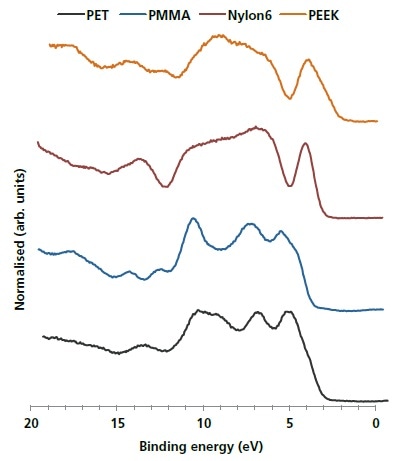
He II excited UPS valence band spectra of four common polymers.
Versatile ESCApe Software for Acquisition and Processing
The ESCApe software has been created to ensure that users’ interaction with the spectrometer is made as easy as possible, combining acquisition and processing to completely exploit the hardware automation. One example of this is the group array analysis capability within the ESCApe software. This feature enables users to drag an area over a sample and define a range of analysis points. Spectra are later acquired from these analysis points.
Automated identification and quantification of peaks enable users to easily produce a color concentration map of detected elements over the surface of the sample.
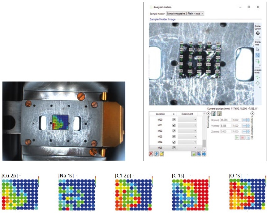
Group array analysis of an inhomogeneous distribution of Cu-nanoparticles on a graphite substrate. The color scale used to represent relative elemental concentration.
Into the Bulk—Ion Sputter Sources
The Minibeam 6 Arn+ gas cluster ion source (GCIS) or the Minibeam 4, high-flux Ar+ monatomic ion source is used to configure the AXIS Supra+. These two ion sources are completely incorporated into the ESCApe acquisition software for depth profile experiments or sputter cleaning. Gas handling is completely automated for sputter profiling and contains pump/purge sequences to enable the transition to helium gas for ion scattering spectroscopy (ISS).
The Minibeam 6 is a type of multi-mode ion source that can work in Ar+/He+ monatomic ion mode or Arn+ cluster mode, as needed. The latest development of gas cluster ion sources has paved the way for depth profiling organic materials with retention of chemistry across the profile. Additionally, depth profiles via intricate multi-layer materials like flexible electronic devices and organic LEDs can now be realized with Arn+ cluster ions.
The Minibeam 6 can produce 20 keV Arn+ cluster ions, and this capability has resulted in the successful etching of inorganic materials like titanium dioxide with considerably less chemical damage caused by the cluster ion etch relative to conventional monatomic ion etching.
- Both sources can operate with He+ ions for optional ISS mode
- Automated introduction of gas and regulation of pressure
- An option of monatomic or multi-mode gas cluster ion sources
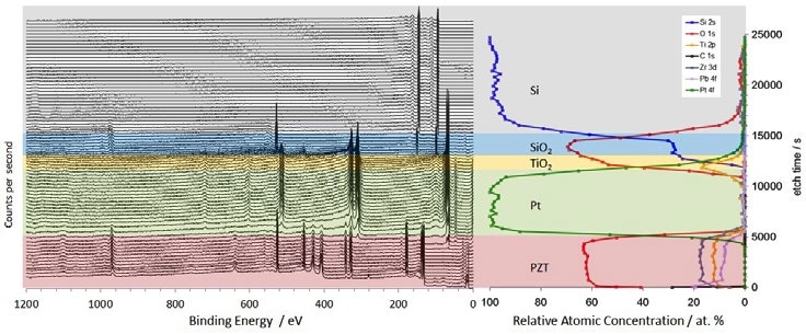
Sputter depth profile survey spectra (left) and elemental concentration as a function of etch time (right) through a PZT/Pt/TiO2/SiO2/Si multilayer sample using 20 keV Ar500+ ions.
Unrivaled Instrument Automation
Sample handling is fully automated in the AXIS Supra+, setting it apart from other spectrometers. The automatic exchange of samples toward the conclusion of an experiment enables continuous operation. Unparalleled levels of automation extend to regular maintenance aspects like computer-controlled bake-out and further degassing of filaments.
In addition, automation includes the monochromator mirror and X-ray source so that calibration and switching between the Al Kα source and the optional Ag Lα excitation source are fully controlled by the computer. The motorized multi-position anode also ensures continued optimized performance.
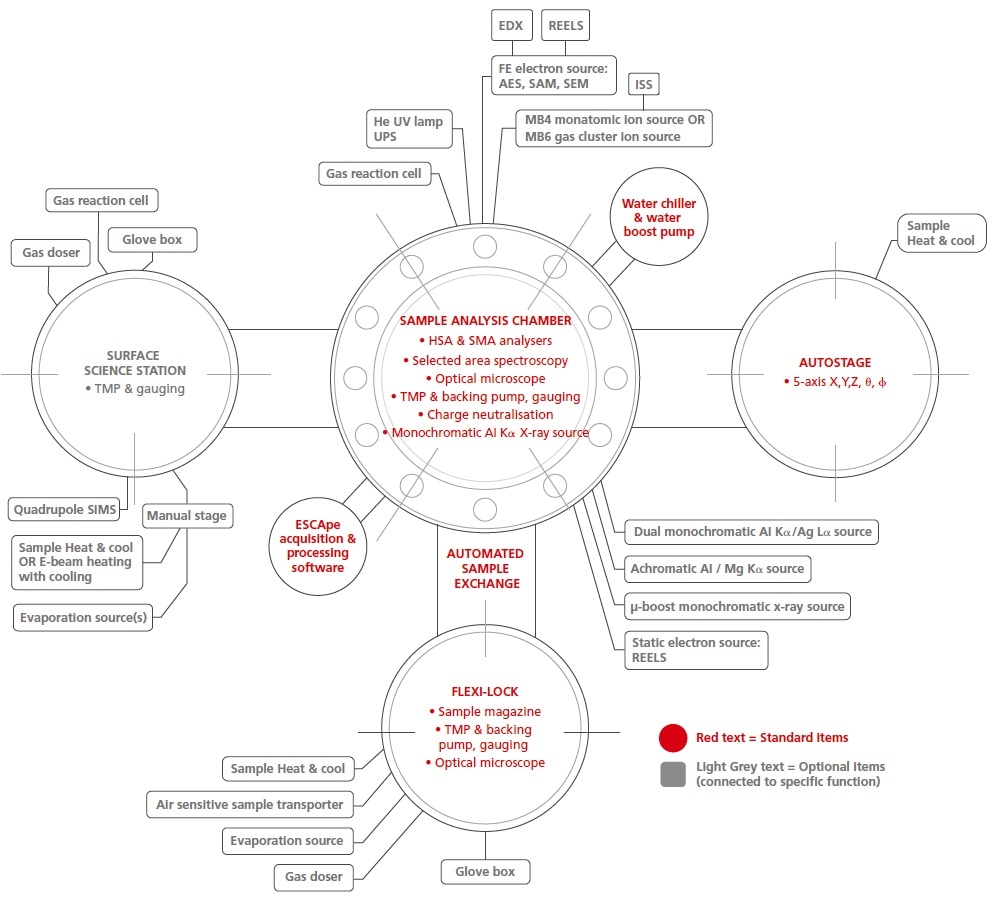
System Configuration Overview
The AXIS Supra+ provides maximum levels of automation together with the flexibility to include complementary sample preparation and/or surface analytical methods. Several optional items described below are provided as upgrades post-installation.
Kratos Analytical has an established track record in developing surface analysis instrumentation hardware. Physicists and mechanical engineers at Kratos Analytical can offer solutions for surface modification, sample handling, and further technique integration for the AXIS Supra+.
AXIS Supra photoelectron spectrometer