Xradia, Inc. announced today that several of its customers have used VersaXRM 3D X-ray microscopes to achieve ground-breaking research results not previously possible in their labs. The VersaXRM family has introduced unprecedented resolution for 3D non-destructive imaging at high contrast into laboratory environments, enabling synchrotron-quality research for a broad range of applications. [See related announcement: "Xradia Expands VersaXRM Family"]
 Xradia VersaXRM Facilitating Studies in Carbon Sequestration
From Synchrotron-to-Lab
Xradia VersaXRM solutions have enabled research once achievable only at Synchrotron beam-lines with selective user access.
According to Dr. Dominique Bernard, CNRS Senior Researcher, Ferroelectric Materials, Ceramics and Composites, Institut de Chimie de la Matière Condensée de Bordeaux (ICMCB), "When evaluating the VersaXRM, it quickly became clear to us that we were seeing results comparable and in many cases exceeding those previously seen in our Synchrotron studies. Not just in resolution, but also in contrast. We have been using the VersaXRM for five months now, and are pushing the instrument to the limits. We have already imaged a very wide range of samples from prehistoric fossils to complex structured multi materials. We are achieving feature detectability down to 70nm and are looking forward to seeing where this technology will take our science in the future."
Additional highlights include recent studies that take a closer look at the structure of natural energy resources and composite materials such as those used in the aerospace industry:
ENERGY
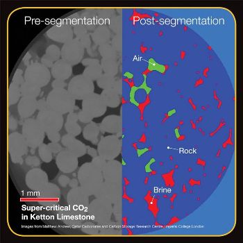
Critical applications leveraging non-destructive X-ray imaging and the VersaXRM family include developing and understanding new climate mitigation techniques, such as carbon sequestration (storing carbon dioxide underground to prevent its escape to the atmosphere). Imperial College, London used the VersaXRM-500 with special sample holders that simulate underground pressures and temperature for in situ studies of carbon storage processes, enabling them to observe a realistic representation of how the carbon dioxide is trapped.
"The use of the VersaXRM-500 has been a big success already and we have achieved very high quality, supercritical images of CO2," says Martin Blunt, Professor of Petroleum Engineering at Imperial College. "These are the highest resolution, highest contrast 3D images ever captured in situ at realistic high pressure fluid flow conditions. These images confirm that CO2 can be trapped in the pore space of the rock, which helps in the design of secure carbon dioxide storage as part of our efforts to prevent global climate change."
MATERIALS SCIENCE
In materials science, understanding and quantifying how materials behave under external environmental conditions (load, temperature, bias, humidity, etc.) ranks among the central goals for quickly and efficiently predicting performance and designing advanced materials for tomorrow's applications. This over-arching mission was recently reflected in the U.S.-based Materials Genome Initiative. At the University of California, Irvine, Professor Lizhi Sun is using the VersaXRM-410 to nondestructively characterize the microstructure and mechanics of composite materials with applications in civil, mechanical, aerospace and biomedical engineering.
"The newly installed VersaXRM-410 lets us characterize the behavior and local deformation of materials in 3D in their native environments (in situ) while uniquely maintaining sub-micron resolution across an array of sample dimensions and environments," states Professor Sun. "What's even more powerful is that we can extend the understanding of a material's microstructure to the 4th dimension (3D plus time), by studying how a microstructure evolves over time and quantify that change. Only non-destructive X-ray tomography lets us achieve that goal."
VersaXRM has also recently advanced studies that aid the invention of stronger and more weather-resistant building materials, three-dimensional mapping of neurons in the brain, and pioneering a new method for petroleum rock analysis to determine the most efficient natural resource extraction process.
The VersaXRM: Extending the Boundaries of Science
Xradia Chief Materials Scientist and Vice President of Marketing, Dr. Kevin P. Fahey, noted, "When we launched the VersaXRM family two years ago, we could only begin to imagine the types of research that would be enabled by its capabilities. Looking forward, XRM stands to become an increasingly prominent component of a distributed, laboratory-based network of microscopes that, alongside complementary analysis techniques and materials synthesis knowledge, will advance research and applications in many areas."
The VersaXRM family and its unique architecture offer the only imaging solutions that combine:
- Non-destructive X-ray imaging, preserving and extending the use of valuable samples
- High resolution: 0.7 to 0.9 μm true spatial resolution with minimum achievable voxel sizes ranging from 70-100 nm*
- High contrast: absorption contrast and unique phase contrast enable unparalleled imaging quality, whether for soft materials such as unstained tissues or low-Z materials like carbon fibers and polymers, with the ability to differentiate between phases of similar density
- RaaD™ Resolution at a Distance: unprecedented resolution for the widest range of sample dimensions, in situ imaging, and interior tomography
- Industry-leading 4D and in situ capabilities for a broad range of sample sizes and types Synchrotron optics in a lab-based solution
*About Resolution
Voxel (sometimes referred to as "nominal resolution" or "detail detectability") is a geometric term that contributes to but does not equal resolution. Xradia specifies on spatial resolution, the most meaningful measurement of an instrument's performance.