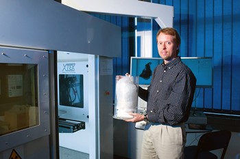
The University of the Witwatersrand in Johannesburg is using a Nikon Metrology micro-CT scanner in its Palaeosciences Centre.
Discovered in South Africa in 1924, the Taung Child was the first hominin fossil to show both human and ape-like characteristics, a species later named Australopithecus africanus. Estimated to be 2.5 million years old, the fossil consists of most of the face, a mandible with teeth and a natural limestone cast of the inside of the cranium (endocast). Anatomical studies of this and similar fossils show that the species regularly walked upright, but the brain was only ape-sized.
Over the years, academics studying the Taung fossil have said that the child, who was estimated to be three to four years old at time of death, had a skull with open sutures that permitted extensive post-natal brain growth. This suggested a larger brain size as an adult than had previously been thought, implying that it was a true ancestor of Homo sapiens (modern humans). More recent studies relying on medical CT (computed tomography) technology reinforced that view.
Now the Palaeosciences Centre of the University of the Witwatersrand (Wits), Johannesburg, is taking another, more in-depth look at the partial cranium and endocast using a Nikon Metrology microfocus X-ray CT (micro-CT) scanner. The idea is to see if the previous research conclusions can be corroborated using the much greater spatial resolution, typically 10 to 100 times that of a medical CT scanner. There are few such non-destructive, radiographic 3D imaging facilities in the world available to institutes studying prehistoric life.
Dr Kristian Carlson, Senior Researcher in the Wits Evolutionary Studies Institute (ESI), said, "The higher resolution surface detail yielded by our Nikon Metrology equipment enabled better, more precise analysis of the partial cranium and curved surfaces of the endocast than had previously been possible.
"Prof Ralph Holloway of Columbia University, Prof Douglas Broadfield of Florida Atlantic University and I have written a paper on the subject that is currently undergoing peer review, which we hope will be published later in the year."
The Taung Child specimen was scanned in the Nikon Metrology XTH 225/320 LC dual source micro-CT system, which uses the manufacturer's Inspect-X software for controlling the machine and for image reconstruction. The scanner can accommodate objects falling within a 23 cm sphere, or longer items if sections are scanned and data stitched together. For maximum operational flexibility, the machine is equipped with both rotating and static reflection 225 kV targets and a more powerful 320 kV static target to enable denser fossil and geological samples to be penetrated. Visualisation and analysis software packages used are VG Studio Max from Volume Graphics and Avizo Standard and Avizo Fire from FEI Visualization Sciences Group.
The flexibility of being able to equip the system with different sources together with the presence of a very experienced local service and support team were two of the main reasons that the research centre opted for Nikon Metrology as a supplier.
To obtain a detailed 3D image of the partial cranium, the X-ray source was set at 135 kV and 400µA, with 6,000 projections created of the rotating specimen. The magnification of the projected fossil image on the panel resulted in an isotropic voxel (3D pixel) size of 55.5 µm, giving very fine spatial resolution. Automated protocols for correcting beam hardening and reducing noise were applied.
Non-destructive analysis is the key to research of precious specimens
Fragile, rare and often valuable specimens can be investigated without damaging them using CT imaging. Rather like MRI scanners in hospitals that use radio waves to scan a patient, CT takes X-ray cross sections through a specimen and reconstructs them digitally to produce a virtual 3D computer model that can be manipulated, sectioned, dissected and measured, internally as well as externally. A major advantage of CT is that it can separate materials based on their density characteristics, so hardened sediments can be easily separated from fossilised bone in the 3D visualisation.
Another useful application is the ability to reconstruct broken bones virtually and to mirror-image a right-hand bone to simulate what the left would look like to build a more accurate picture. Additionally, if a decision is taken to physically dissect a specimen, it can be done faster, more accurately and more safely by running a CT scan first to identify the location and orientation of a fossil within a rock.
This type of research can be carried out by scientists in many other disciplines and the data shared with colleagues all over the world. Already the micro-CT facility in Johannesburg is being used by other researchers within Wits involved in botanical sciences, medical research, geology, mining and archaeology, as well as by the university's School of Art.
Collaboration extends beyond the university, with people from four or five museums and a similar number of academic institutions in South Africa taking advantage of the equipment. The Karoo region of the country is yielding dinosaurs and even more ancient reptiles and amphibians for analysis.
Prof Bruce Rubidge, Director of the Evolutionary Studies Institute at Wits, commented, "One of the goals in purchasing this equipment was to build human capacity and expertise amongst southern African scientists and to lead virtual-based palaeontological studies, rather than having this work undertaken by overseas specialists.
"We are excited to see this goal being realised as more scientists and students utilise the equipment and learn the necessary skills in the process."
CT scanning opens new possibilities for 3D printing
Having acquired X-ray slices through a specimen to produce digital files, it is an easy step to translate the resultant CT datasets into STL (stereolithographic) files to feed into a 3D printing machine for creating a physical model of a fossil, even if it is still encapsulated inside a rock. The value of this approach was recognised at the outset by the team at Wits, which also bought a Z-Printer 450 additive manufacturing machine from Z-Corp to build models layer by layer from powder.
A slightly textured surface is important when recreating prehistoric specimens, as shiny surfaces would look unnatural. The powder layers, which are down to 87.5 microns thick, are ideal for imparting the right appearance. Colour can be introduced, giving variety to displays in the Palaeosciences Centre. Models can be scaled up or down to assist in education or research.
A direct 1:1 facsimile of the specimen can be printed from which a mould can be made to produce accurate casts of particularly delicate specimens. STL file sizes are smaller than the 3D volume from which they were made and so they can be conveniently emailed or embedded in technical papers.
Conclusion
Micro-CT is a powerful, non-destructive imaging technique for visualising 3D objects, both externally and internally, allowing rich detail to be revealed without any damage to the specimens. Digital preparation can be quicker than many mechanical or chemical preparations. It also offers the flexibility to revise the virtual preparation or try different potential outcomes, whereas physical preparation is permanent. Original CT data can be stored with the actual specimens to provide a permanent digital record, with the option of repeating the imaging process in the future as technology advances.
Eventually, entire collections of museums and universities around the world could be available in digital form over the web to scientists and the public. The Natural History Museum in London, which also owns a micro-CT scanner, contains approximately 75 million specimens that will take generations to digitise to reveal hitherto hidden secrets. It is particularly fitting that Wits also owns a high performance CT scanner, as the university is the custodian of one of the largest collections of fossils in the southern hemisphere.
All Nikon Metrology X-ray machines are built at the group's factory in Tring, Hertfordshire, UK. An even more powerful 450 kV micro-CT scanner is also manufactured there. The high energy source is capable of investigating larger or denser samples and can therefore open new avenues of research in the palaeosciences, and specifically in palaeontology.