Jun 4 2014
Anyone involved in macromolecular crystallography will know that for many years scientists have had to rely on a multi-stage process utilizing protein, usually expressed in engineered cells, which is then extracted and purified before crystallization in vitro and finally prepared for analysis.
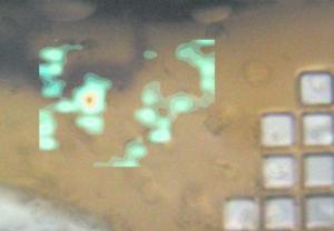 This shows the results of a grid scan from in cellulo data collection shown as a contour plot of the DISTL output 'good Bragg candidates.' Credit: Axford et al
As a counter to this time-consuming and substantial scientific effort, there are a number of examples of protein crystallization events occurring in vivo, with next to no human input. In a case presented in a recent paper an insect virus exploits the phenomenon as part of its life cycle. Not surprisingly an issue with intracellular protein crystals is that they are typically very small, limited by the size of the cell. However, microfocus beamlines at synchrotron light sources prove here to be capable and refined in the analysis of micron-scale in vivo samples.
A group of scientists from the Diamond Light Source and the University of Oxford, UK [Axford et al. (2014), Acta Cryst. D70, 1435-1441; doi:10.1107/S1399004714004714] has been able to study crystals inside the cells directly using X-ray analysis without complex attempts to extract and prepare samples. It would not be out of place to assume that the presence of cellular material might compromise the experiment. However, the researchers' results show that the exact opposite may actually be true; the cell maintains the crystals in an environment amenable to the collection of data.
It will be interesting to see if an improved understanding of protein crystallization in vivo can bring more targets within reach of such analysis. Certainly continued technical developments, including increased photon flux and reduced beam size, will improve the signal-to-noise ratio. Together with more efficient data processing, this means that we will be able to do more with less and exploit novel microcrystal targets of increasing complexity for in vivo structural studies.
Source: http://www.iucr.org/