Sep 22 2014
A new technique developed by researchers at Xi’an Jiaotong University and Forschungszentrum Jülich, the Ernst Ruska-Centre for Microscopy and Spectroscopy with Electrons (ER-C), allows the reconstruction of crystal structures with atomic-scale accuracy in three dimensions.
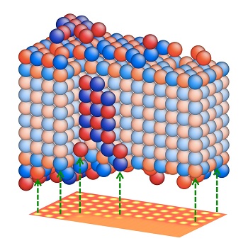 From the electron microscope image of nanocrystals – here a magnesium oxide nanocrystal (below) – the three-dimensional structure is reconstructed with atomic precision using a new method developed by researchers from Jülich and Xi’an. The red spheres represent magnesium, the blue, oxygen. (Source: Forschungszentrum Jülich)
The shape and surface texture of nanomaterials greatly influences their physical and technical properties. As a result, material scientists and physicists are very interested in determining a nanomaterial’s structure with atomic precision from all angles.
The team of researchers carried out a series of tests from different angles and successfully computed the spatial arrangement of the atoms using a single ultra-high resolution electron microscopic image.
The new method is ideal for performing the spatial mapping of samples which are highly sensitive to high-energy measurement beams. The new 3D measuring technique’s relatively short data acquisition time holds potential for monitoring a chemical reaction’s transient intermediate steps and enables both heavy and light chemical elements to be detected.
In this study, a magnesium oxide sample was placed in a microscope in such a manner that the atoms at the crystal lattice intersections lay over each other, thus creating atom columns across the observation axes. The signal-to-noise ratio was improved using a special imaging mode, which allowed the observation of subtle variations. As a result, the researchers were able to locate individual atoms in the columns along the direction of the beam.
The research team then compared the image to computer calculations of the spatial structure. And began matching up the model crystal step-by-step, until the reconstructed image matched the electron microscopic image. The researchers verified the uniqueness of the study results by conducting extensive statistical tests, which revealed that the technique not only has atomic-scale sensitivity, it can also distinguish between the oxygen and magnesium of the thin crystalline sample.
This study was published in the Nature Materials journal.