Kwansei Gakuin University is situated in the Hyogo prefecture in the Japanese cities of Nishinomiya and Sanda.
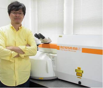 Professor Noboru Ohtani of the Department of Nanotechnology for Sustainable Energy, School of Science and Technology at Kwansei Gakuin University with his Renishaw inVia Raman Microscope.
Professor Noboru Ohtani is a member of the Department of Nanotechnology for Sustainable Energy in the School of Science and Technology. His major fields of research are the study of wide band gap semiconductors and crystallographic defects.
Crystallographic defects in 4H-SiC epitaxial wafers, such as dislocations and stacking faults, limit the commercialisation of SiC devices and must therefore be eliminated or reduced to levels lower than some critical density. The department’s research goal is to establish SiC crystal growth processes that can produce large, ultra-high quality SiC epitaxial wafers. To achieve this, they try to clarify the cause and formation mechanism of crystallographic defects in SiC bulk crystal and epitaxial film.
Crystallographic defects give rise to residual stresses in the crystals. The stresses can occur through a variety of mechanisms in 4H-SiC crystals. For example, temperature gradients in the grown crystals, which are a primary driving force for crystal growth, lead to plastic deformation of the crystals during the growth and/or cooling process. This deformation results in residual stresses when the crystals are cooled to room temperature.
The spatial variation of stresses in the crystals can be measured using Renishaw’s inVia confocal Raman microscope. These measurements provide valuable information about the formation of defects during physical vapour transport (PVT) growth and chemical vapour deposition (CVD) processes. This information is used to improve the crystal growth process.
Professor Ohtani’s laboratory also uses high resolution X-ray diffraction (HRXRD) to characterise stress distribution. Raman microscopy provides complementary information to the HRXRD data but with much higher spatial resolution. When asked why the inVia was chosen for the lab, Professor Ohtani said: “The key benefit is the ultra-high speed data acquisition system, which results in a higher sensitivity to measuring stresses in the materials compared to other Raman systems.”
Professor Ohtani and his colleagues recently published a paper with the Materials Science Forum describing the ‘structural and electrical characterization of the initial stage of physical vapour transport growth of 4H-SiC crystals.’1 It illustrates the power of micro Raman imaging to help show the influence of heavily-doped nitrogen donors on the defect formation in SiC crystals.
Please visit www.renishaw.com/invia for further details of Renishaw’s inVia confocal Raman microscope.
Reference
Structural and electrical characterization of the initial stage of physical vapour transport growth of 4H-SiC crystals, Materials Science Forum Vols 821-823 (2015) pp 90-95. www.scientific.net/MSF.821-823.90 T Takahashi et al.
About Renishaw
Renishaw is a global company with core skills in measurement, motion control, additive manufacturing and precision machining. The company also specialises in the production of high quality optical spectroscopy products and is a recognised leader in Raman spectroscopy. The team of scientists and engineers within Renishaw’s Spectroscopy Products Division specialize in the development, application and production of high performance, configurable Raman spectrometers.