May 5 2016
Researchers at the Johns Hopkins University report that each year, birth defects, surgery, or trauma have resulted in an estimated number of 200,000 people requiring bone replacement surgery in the face or head.
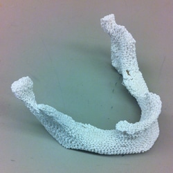 A sample 3-D printed scaffold that matches the lower jaw of a female patient. (Credit: Johns Hopkins Medicine)
A good framework to fill in the missing bone can be developed by combining 30% of pulverized natural bone with a unique artificial plastic and producing the required shape with a 3D printer. According to the Johns Hopkins research team, this is an effective recipe for treating such medical conditions. A mice study has also shown that a blend of artificial and natural materials works best. The results of the study have been reported in a paper published online in ACS Biomaterials Science & Engineering.
In a traditional treatment, surgeons take out part of a patient’s fibula, cut it into the required general shape, and then implant it in the exact location. According to Warren Grayson, Ph.D., the report’s senior author and associate professor of biomedical engineering at the Johns Hopkins University School of Medicine, the surgical procedure causes leg trauma and is not an optimized solution, because it is not possible to shape the straight fibula and fit it in the slight curves of the face. As a result, researchers turned to 3D printing or additive manufacturing that produces 3D objects from a digital PC file by depositing successive, ultrathin material layers.
This process effectively creates highly accurate structures, including anatomically precise structures from plastic.
[But] cells placed on plastic scaffolds need some instructional cues to become bone cells. The ideal scaffold is another piece of bone, but natural bones can’t usually be reshaped very precisely.
Warren Grayson, Ph.D., Associate Professor of Biomedial Engineering, School of Medicine, John Hopkins University
To perform the experiments, the researchers initially developed a composite material that would integrate the printability and strength of plastic with the biological data present in the natural bone. They started with biodegradable polyester called polycaprolactone (PCL); PCL is widely used for making polyurethane, which is an FDA-approved plastic and is also used in other clinical applications.
PCL melts at 80 to 100 degrees Celsius (176 to 212 Fahrenheit) — a lot lower than most plastics — so it’s a good one to mix with biological materials that can be damaged at higher temperatures.
Ethan Nyberg, Graduate Student, John Hopkins University
As previous studies have shown, PCL does not support the formation of new bones very well, despite it's excellent strength. Due to this the researchers mixed the PCL with increasing quantities of bone powder, which was made by crushing the porous bone of cow knees after removing the cells from it.
Bone powder contains structural proteins native to the body plus pro-bone growth factors that help immature stem cells mature into bone cells. It also adds roughness to the PCL, which helps the cells grip and reinforces the message of the growth factors.
Warren Grayson, Ph.D., Associate Professor of Biomedial Engineering, School of Medicine, John Hopkins University
Grayson added that printability was the initial test for the composite materials. While 5%, 30% and 70% blends of bone powder performed well, 85% bone powder had very little PCL glue to preserve distinct lattice shapes, and was discarded from future experiments.
It was like a chocolate chip cookie with too many chocolate chips.
Ethan Nyberg, Graduate Student, John Hopkins University
In order to determine whether the scaffolds promote the formation of new bone, the team added human fat-derived stem cells - taken during a liposuction procedure - to scaffolds kept in a nutritional broth without any pro-bone ingredient. After a period of three weeks, it was observed that cells grown on 70% bone powder scaffolds, displayed gene activity that was many times higher in three genes, suggesting the formation of bones as opposed to cells grown on pure PCL scaffolds alone. Cells grown on 30% bone powder scaffolds displayed large yet a less remarkable increase in the same genes.
When the researchers introduced the beta-glycerophosphate to the cells’ broth to allow their enzymes to deposit calcium, the cells grown on 30% scaffolds generated about 30% more calcium for each cell, while those on grown on 70% scaffolds generated more than twice as much calcium for each cell, compared to cells grown on pure PCL scaffolds.
To further test their scaffolds, the researchers used mice that had extremely large holes in their skull bones. It was seen that the bone wounds were relatively large to heal without any form of intervention. Mice, which received scaffold implants infused with stem cells, experienced bone growth inside the hole over the 12-week period of the experiment. CT scans revealed that bone grew as much as 50% in scaffolds that had 70% or 30% bone powder, as opposed to those with pure PCL.
In the broth experiments, the 70 percent scaffold encouraged bone formation much better than the 30 percent scaffold, but the 30 percent scaffold is stronger. Since there wasn’t a difference between the two scaffolds in healing the mouse skulls, we are investigating further to figure out which blend is best overall.
Warren Grayson, Ph.D., Associate Professor of Biomedial Engineering, School of Medicine, John Hopkins University
While the use of decellularized cow bone has been approved by the FDA for clinical use, the team is hoping to experiment with bone powder produced from human bone, as it is more extensively utilized clinically. They also intend to test the interior designs of the scaffolds to make it more natural and less geometric, as well as additives that promote the growth of new blood vessels to penetrate the scaffolds, which will be required to extend the life of denser bone implants.
Other co-authors of the report are Ben Hung, Miguel Dias, Bilal Naved, Christina Holmes, Amir Dorafshar, and Jennifer Elisseeff of the Johns Hopkins University School of Medicine.
The study was funded by the National Institute of Dental and Craniofacial Research (F31 DE024922), the Russell Military Scholar Award, the Department of Defense, the Maryland Stem Cell Research Fund and the American Maxillofacial Surgery Society Research Grant Award.