Jun 9 2016
The Sol M. Gruner (SMG) detector group at Cornell University has created and tested a novel imaging electron detector that enables quick and accurate measurements of magnetic and electric fields at the atomic level. The new detector can record an image frame in 1/1000 of a second and identify from 1 to 1,000,000 electrons per pixel. It is 1000 times more powerful and 100 times faster than standard electron microscope image sensors.
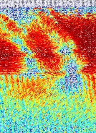 This image might look like an impressionist sunset, but it is actually a vector map of the measured deflections of an atomic-sized electron beam scanned across different polar domains in the ferroelectric bismuth ferrite. The image was recorded in about a minute by the new electron microscope pixel array detector. (Image courtesy of Cornell University)
The Impact
The new imaging electron detector captures the entire transmitted electrons and helps measure materials properties quantitatively, including internal magnetic and electric fields, which are vital for use in electronics and memory applications.
Summary
Scientists at Cornell University created and tested a new electron microscope detector that can quantitatively measure magnetic and electric fields from micrometers to atomic resolution. The imaging device is based on a solid-state x-ray detector technology that was developed by the scientists over the past fifteen years and now improved to operate as a high dynamic range, high-speed electron diffraction camera.
The dynamic range indicates the highest signal range that can be identified by a pixel and the ensuing electron microscope pixel array detector captures the image frames in less than a millisecond and detects primary electrons from 1 to 1,000,000 pixels per image frame. This is 1000 times more powerful and 100 times faster than standard electron microscope image sensors. These material properties help the researchers to record the whole unsaturated diffraction patterns in scanning mode, capture dark field, bright field and phase contrast details simultaneously, and investigate the scattering distribution thoroughly, thus paving the way for novel multichannel imaging modes. The researchers can extract polarity, tilts and local strains, as well as magnetic and electric fields based on the analysis of the spatially resolved diffraction patterns.
Funding
Pixel array detector (PAD) development in SMG’s lab is supported by the U.S. Department of Energy (DOE), Office of Science (SC), grant DE-FG02-10ER46693 and by the Cornell High Energy Synchrotron Source (CHESS), which is supported by the National Science Foundation (NSF) and the National Institutes of Health (NIH) National Institute of General Medical Sciences via grant DMR-1332208. The PAD architecture used was developed as an x-ray detector over the last decade and a half as a collaboration between SMG’s group and Area Detector Systems Corp. (Poway, CA) under NIH grant R44 RR014613 and DOE/SC grant DE-FG02-97ER62443.
Cornell for Nanoscale Science’s Kavli Institute supported the x-ray PAD adaptation to the scanning transmission electron microscope (STEM). The Cornell Center for Materials Research, an NSF Materials Research Science and Engineering Center (MRSEC), supported the Electron microscope data acquisition (KXN, DAM) under the DMR-1120296 grant. The base annular dark field detector case for the device was provided by Fischione Instruments’ Paul Fischione.