Aug 2 2016
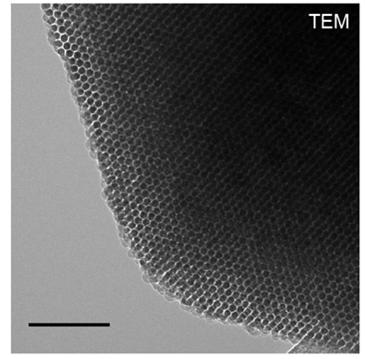 A transmission electron microscope image taken at Argonne shows the honeycomb structure of the silicon nanowires. (Credit: Jiang et al.)
Silicon particles that could lead to the development of “biointerface” devices have been engineered by a group of scientists from the University of Illinois at Chicago, the U.S. Department of Energy's (DOE's) Argonne National Laboratory, the University of Chicago, and Northwestern University.
The silicon particles measure one-fiftieth the width of a single strand of human hair, and can help create biointerfaces to make heart cells beat and nerve cells fire.
The head of the University of Chicago team, Bozhi Tian, stated that the deformable nature and the ability to produce a local electrical effect makes these particles capable of establishing unusual biointerfaces on cell membranes.
Biological systems are soft, and if you want to design a device that can target those tissues or organs, you should match their mechanical interface as well. Most of the current implants are rigid, and that's one of the reasons they can cause inflammation.
Bozhi Tian, University of Chicago
Unlike biointerfaces made of materials such as carbon and gold, the biointerfaces created by these particles will degenerate with time, said Yuanwen Jiang, co-author of the research and a graduate student in Tian’s team. As a result, a second operation of the patient to remove the particle can be prevented.
Tian and Jiang believe that the material, employed with light, could be useful to excite a variety of cells and has numerous potential applications in the biomedical field. The process of nano-casting was used to develop the mesostructured silicon. The material earned the name due to the complex internal structure made of nanoscopic wires.
Argonne News Brief: Silicon particles use light to activate cells
Synthetic silicon dioxide was used for its beehive-like structure, to produce the particles that measure between one to five micrometers in size. Just as a blacksmith pours molten metal in a cast iron mold, the scientists poured semiconductive silicon into the beehive-like structure. A bundle of wires that were connected by thin bridges were obtained, after the external structure was removed using acid.
A sample of the particles was administered to cultured rat dorsal root ganglia neurons, present in the peripheral nervous system, to see if the particles can impact cell behavior. Using light to heat the silicon particles, the researchers succeeded in activating the neurons, which allowed electricity to pass through the cells.
Unlike traditional biointerfaces that require a connection to a source of energy, silicon particle require only light, making the system completely wireless. The particles can be activated through the skin after being directly injected into the right place.
Neuromodulation could take full advantage of this material, including its optical, mechanical and thermal properties.
Yuanwen Jiang, Graduate Student, University of Chicago
Jiang said that in addition to impacting neurodegenerative disorders by controlling neurons, Tian’s team has also used the material to manipulate the beating of heart cells.
Resources from the Center for Nanoscale Materials, a DOE Office of Science User Facility, and Argonne X-ray Science and Chemical Sciences and Engineering Divisions have been used for the study.
The 32-ID and 12-ID-B beamlines from another DOE Office of Science User Facility, Advanced Photon Source, were used by the researchers to take transmission X-ray microscopy nano-computed tomography of the samples, to take X-ray scattering measurements, for transmission electron microscopy, and scanning electron microscopy. Focused ion beam lithography expertise and instruments, and tools to design optical masks were provided by the Center for Nanoscale Materials.
The study titled ‘Heterogeneous silicon mesostructures for lipid-supported bioelectric interfaces’ was published on the June 27 issue of Nature Materials. Il Woong Jung, Di-Jia Liu, Xiaobing Zuo, Vincent De Andrade, and Xianghui Xiao were the Argonne co-authors of the study.
The research was financially supported by the National Science Foundation, the University of Chicago Start-up Fund, the Air Force Office of Scientific Research, the Searle Scholars Foundation and the National Institutes of Health.