A key to understanding the function of cells and tissues is the ability to image their structure. The way that musculoskeletal tissues are structured, for example, will determine their ability to perform their mechanical functions. In cellular material, the cell membrane mediates the exchange of material with its environment, helping to drive biological responses.
Accurately Obtaining Microscopic Images of Biological Samples
Many biological specimens, however, present obstacles to accurate microscopic imaging. Many of these materials are non-conducting, making for a challenge in obtaining a stable, high resolution image. Their organic composition also make them susceptible to shrinkage under the influence of charged particle beams. Thus it is desirable to create minimum impact to these sensitive structures in order to image them successfully.
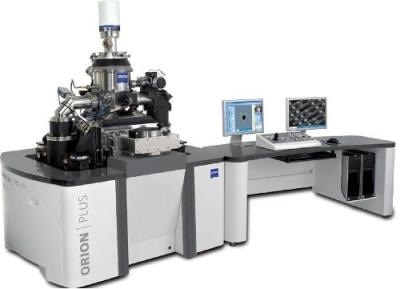
Imaging Biological Samples Using The ORION PLUS Helium Ion Microscope
We can take advantage of the unique beam-sample interaction of a primary helium ion beam in order to get past the challenges described above. For samples that charge it is often possible to simply image at lower beam current in order to allow charge to dissipate and to create a stable image. The consistent nature of the charge flow in an ion microscope (positive charge in and negative charge out under all conditions) obviates the often difficult task of searching for the charge equalizing beam voltage as is required in SEM. The ORION® PLUS also can maintain this charge flow balance at high beam energy, bypassing the trade-off between charge control and resolution necessary in SEM.
An example of this is seen in imaging of collagen fibers from the knee joint of a mouse, as seen in the first image below. This sample, prepared by critical point drying, was found to be challenging to image in SEM due to charge related instabilities. In the ORION® PLUS it was possible to obtain a stable image, thus more easily revealing detail on the banding in these fibers. There is also charge neutralization capability available from a low energy electron flood gun.
An example of non-damaging image acquisition is found in the investigation of CHO (Chinese Hamster Ovary) cells. CHO cell lines are very common and important, being utilized for example in drug discovery. The structure of the membranes and of the finger-like structures, called filopodia, on these cells is related to their biology – and thus to their behavior in pharmaceutical research. The ORION® PLUS microscope has shown the ability, as seen in the second image below of critical point dried cells, to provide superior image resolution and contrast. It is also notable that it is possible to image the filopodia at high magnification without shrinking them, which is a problem in SEM imaging.
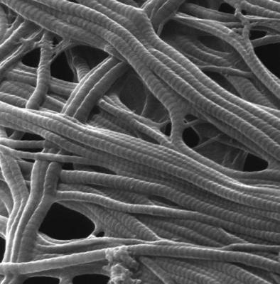
Figure 1. Collagen fibers Imaging in collaboration with Dr. Claus Burkhardt, NMI (Stuttgart, Germany)
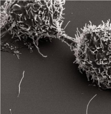
Figure 2. Chinese Hamster Ovary (CHO) cells Imaging in collaboration with Dr. Claus Burkhardt, NMI (Stuttgart, Germany)
The ORION PLUS Capabilitites
Material contrast, non-destructive imaging, charge control
Application
Imaging three dimensional biological structures

This information has been sourced, reviewed and adapted from materials provided by Carl Zeiss Microscopy GmbH.
For more information on this source, please visit Carl Zeiss Microscopy GmbH.