Over the past few decades, light microscopy has become an important and widely used tool in forensic science research. It is a fast and accessible method, and is non-destructive, which is useful when dealing with precious and irreplaceable samples.
Often used in forensic laboratories, specialists use this technique as a first-line analysis for research and investigation, prior to subsequent techniques. In forensic research, other specialized forms of microscopy such as comparison microscopy and polarized light microscopy are also utilized.
The forensic science research team at the Abertay University in Scotland has used Evident opto-digital light microscopes to expedite a number of research projects. Opto-digital technology integrates the latest in digital and optical technologies and allows fast and comprehensive analysis and measurement in materials science applications.
The Evident DSX range covers measurement, investigation, and reporting, and allows intuitive operation. This means in-depth microscopy knowledge is no longer required for precise inspection.
In addition, the DSX series include digital capture and display which allows on-screen viewing for long-term training, discussions or operation. The capabilities of DSX series opto-digital microscopes allow the research team to analyze different types of evidence in more detail, including pen marks, and fingerprints.
Revealing Latent Fingerprints
Fingerprints are unique to each individual. They are left behind on contact with a surface, which serves as a critical source of identification. Fingerprints are composed of skin ridges, and each skin ridge has a line of pores through which sweat is released, as shown in Figure 1.
Most often, fingerprints are formed by the sweat, rather than by physical imprints or other residue. Such prints need additional treatments to be visualized, depending on the surface on which they were left.
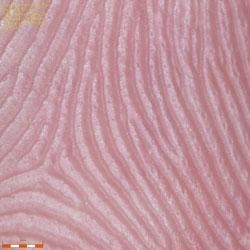
Figure 1. Fingerprint pores
One specific area of research is the retrieval of latent prints from metal surfaces where the original residue of the print has been cleaned with a cloth. However, as soon as a contact is made, the sweat contains chemicals that react with the metal and the fingerprint is imprinted onto the surface, leaving an persistent mark.
Abertay University researchers are exploring ways to view these marks by heating the metal over 400ºC, then capturing and studying the images of the mark through the Evident DSX100 opto-digital microscope.
To that end, fingerprints on a wide range of metals were analyzed, along with how their removal using different fabrics, such as cotton, wool and nylon, affected the latent print (Figure 2). Visualization of the print at a high magnification was vital for thorough analysis. This was facilitated by the image stitching function of the DSX100 microscope.
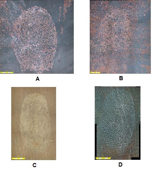
Figure 2. A. Copper wiped with wool; B: Copper wiped with nylon; C: Brass wiped with wool; and D: Brass wiped with cotton.
However, when prints were viewed directly from above, important features could not be distinguished properly. Here, the ability to adjust the head of the DSX100 microscope proved to be particularly useful, enabling the sample to be illuminated and visualized at an angle. Such lighting casts shadows to underline the surface topography for in-depth visualization of the valleys printed onto the metal (Figure 3).
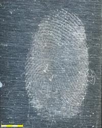
Figure 3. Highlighting fingerprint patterns on metal with oblique lighting.
Revealing Latent Fingerprints from Metal
When fingerprints are wiped from metal, they appear again when the metal is heated to 400°C and beyond. Using the DSX100 microscope, the researchers were able to view latent fingerprints on copper wiped with wool (A) or nylon (B), and on brass wiped with wool (C) or cotton (D), as shown in Figure 2.
Logos on Seized Illicit Pharmaceutical Tablets
At Abertay University, the forensic research team works with Police Scotland, who provide the necessary details to ensure that new research provides the most impact for cutting-edge forensics applications.
Under this collaboration, the DSX100 microscope was employed to examine a set of illicit drugs. Drug suppliers often use alternative substances in place of psychoactive chemicals, and the level of such substances can differ considerably. Hence, it is important to differentiate substances for intelligence purposes.
The Abertay research team is exploring the possibility of doing this by utilizing a combination of physical and chemical properties. The aim is to develop a statistical model that could help detect different populations of tablets in the illegal supply chain, thereby allowing the police to determine links between different seizures and track certain batches of drugs back to their supplier.
The Evident DSX100A microscope was used to assess a batch of seized tablets. Visual characterization was accomplished based on several features, such as design and colour. Most of these tablets included logos, while some even contained defects on the edges.
Again, the tilt angle function of the DSX100 microscope helped in viewing the logos and the edges. Digital capability has enhanced the visualization of these tablets, with what is called as the Extended Focal Image (EFI) tool. Using 3D imaging, EFI conforms to a range of 2D images at different points across the z-axis, creating a composite image across the complete sample and enabling the complete tablet surface to be viewed in detail.
Pen Marks on Paper
Documents may be forged or illegally manipulated for several reasons. Incidents like benefit fraud and identity theft are very common. However, the principle aim of any analytical process is to collect data without damaging or changing the document. Here, the non-destructive nature of light microscopy proves extremely useful. While analyzing such documents, it is important to look at the paper type, ink, and handwriting.
One particular project at Abertay involves distinguishing the ball point pen mark indentations on paper. For instance, when a line is drawn on paper, the pointed tip of the pen alters the paper’s fibres, leaving a mark. The shape of the indentation can differ depending on the pen, the user's style of writing, as well as the quality of the paper.
Other techniques can also be utilized alongside light microscopy to obtain complementary data, like the chemical composition of the ink. Chromatography, however, is limited with gel pen inks, which are not easy to dissolve, so these pens require an alternative means of analysis.
For this project, opto-digital microscopy allowed the required magnifications, which helped in observing the actual fibres of the paper, whilst retaining the colour distribution and range of ink. Also, the indentation depth could also be determined and viewed in the form of a height map. This map can then be visualized and examined in 2D or 3D.
Analyzing Pen Indentations on Paper
Analysis of pen indentations on paper can provide a better understanding about the possible identity of the author. Here, pen marks have been viewed using the DSX100 microscope in brightfield (A), 2D imaging based on a height map (B), and 3D imaging based on a height map(C).
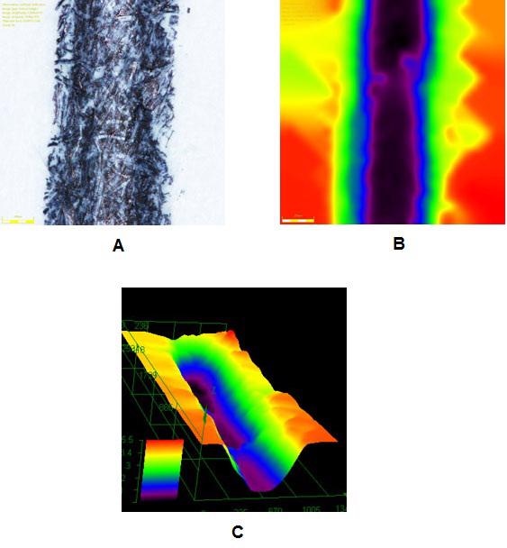
Figure 4. A: DSX100 in brightfield, B: 2D imaging based on a height, and C: 3D imaging based on a height map.
In the Courtroom
The insights gained through light microscopy can also be useful in courtrooms. Information captured in an image can be more easily elucidated to a jury, rather than through a verbal or numerical explanation.
Figure 5 depicts a case of how information can be clearly communicated using the reporting functions of the DSX100 microscope.
In all the three projects illustrated above, the image stitching function was fundamental to image analysis. Whether analyzing an illicit drug, a fingerprint, or a pen mark on a fraudulent document, large images are critical for presenting evidence to a jury. These images can even be zoomed to distinguish the finest details of the sample to gain a greater insight.
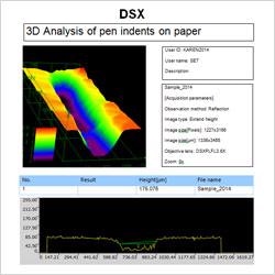
Figure 5. The 3D profile and analysis of a pen indent on paper is reported here alongside additional information such as capture settings.
Conclusion
From the lab to the courtroom, light microscopy offers immense possibilities within the field of forensic science and investigations. The Evident DSX100 opto-digital instrument allowed the forensic research team at Abertay University to gain a better understanding of the wide range of forensic samples.
Light microscopy allows investigators to solve criminal cases and reach a decision faster in a non-destructive manner. This is particularly important in view of budgetary constraints in forensic centers. Thus, light microscopy will continue to improve forensics applications, from research through to applied investigations.

This information has been sourced, reviewed and adapted from materials provided by Evident Corporation - Industrial Microscopy.
For more information on this source, please visit Evident Corporation - Industrial Microscopy