Correlative Raman Imaging and Scanning Electron (RISE) Microscopy (Figure 1) is a unique combination of Confocal Raman and Scanning Electron Microscopy (SEM) Imaging, enabling new possibilities in sample characterization. With this advanced correlative microscopy, it is possible to link ultra-structural surface properties to molecular compound data.
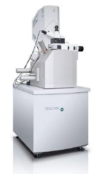
Figure 1. RISE Microscopy
Confocal Raman Imaging
Molecular compounds in a sample can be analyzed using the Confocal Raman Imaging technique, which is a combination of confocal microscopy and Raman spectroscopy. This non-destructive spectroscopic technique is based on the Raman effect caused by the interaction of light with the chemical bonds in the sample. The energy shift in the back scattered light caused by the interaction following vibrations in the chemical bonds can be observed as a unique Raman spectrum.
The Confocal Raman Imaging technique detects and captures the spatial distribution of the chemical components present in a sample. It is also possible to analyze other sample characteristics, including crystallinity, stress and strain states. The information of an entire Raman spectrum can be collected at every image pixel using high-resolution confocal Raman microscopes, yielding a lateral resolution at the diffraction limit of ~200nm. A confocal microscope configuration also has an outstanding depth resolution and the ability to generate 3D Raman images and depth profiles.
Scanning Electron Microscopy
The morphology and topography of a sample can be determined using the SEM technique. This high-resolution imaging technique is compatible with other imaging techniques. It involves scanning of the sample using a focused, high-energy electron beam for generation of images. The electron-sample interactions produce backscattered and secondary electrons, which are detected to yield an image characterizing the morphology, materials orientation, surface structure and topography of the sample.
The SEM technique acquires images at micro- or nanometer resolutions and magnification ranges of 10 – 10.000x. The non-destructive method can also yield a 3D appearance of the surface as it has the large depth of field.
RISE Microscopy
By having the capabilities of both Confocal Raman and SEM Imaging techniques in a single instrument, the RISE Microscopy is able to link ultra-structural surface information to molecular compound data. Sample transfer and repositioning is automatic during switching between the two techniques. It is possible to correlate the acquired results and overlay the images.
The sample’s chemical characteristics can be acquired by using SEM in conjunction with Energy dispersive X-ray spectroscopy (EDX). However, confocal Raman imaging is advantageous over EDX by enabling detection of chemical elements as well as the chemical compounds and molecules of a sample. It is also possible to image the spatial distribution of the components in a sample.
Application Examples
In a research study, a geological diorite sample was analyzed using TESCAN’s RISE Microscope without staining or fixing the sample. The surface structure of the sample was first analyzed using the SEM microscope function of the RISE Microscope (Figure 2A).
Following the surface structure analysis, the sample was then transferred and positioned within the vacuum chamber of the electron microscope for confocal Raman imaging to record the sample are in Raman imaging mode. A complete Raman spectrum representing the molecular composition was produced at each image pixel.
This was followed by overlaying of the acquired SEM and color-coded Raman images for comparison of the ultra-structure with the molecular components (Figure 2B). The resulting Raman spectra are depicted in Figure 2C, enabling to identify each chemical compound based on its characteristic Raman “fingerprint”.
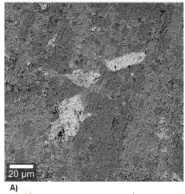
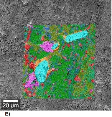
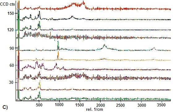
Figure 2. A) SEM Image of a geological diorite sample B) SEM image overlaid with the Raman image. C) The corresponding color-coded Raman spectra display each molecular component of the sample.
In another example, hamster brain tissue placed on a microtome slide was analyzed using the RISE Microscopy. The structural differences that can be observed in the SEM image clearly differentiate the white and gray brain matter. The distribution of the gray and white matter can be more precisely observed when the SEM is overlaid with the color-coded Raman image (Figure 3A). It is possible to clearly differentiate the tissues from their characteristic Raman spectra (Figure 3B).
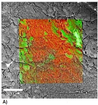
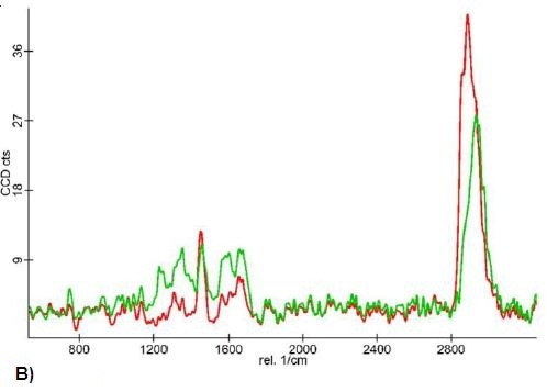
Figure 3. A) Raman-SEM image overlay of a hamster brain tissue sample. B) The corresponding Raman spectra reveal the different spectral characteristics of the white and gray brain matter.
Conclusion
The results clearly demonstrate the ability of RISE Microscopy to link ultra-structural surface data with molecular compound data, opening up new dimensions for in-depth sample characterization. The user-friendliness and outstanding analytic benefits of RISE Microscopy makes it a valuable tool for a myriad of applications, including life science, geo sciences, polymer science, materials science, pharmaceuticals and nanotechnology.
TESCAN Group
Founded in 1991 by a group of managers and engineers from Tesla with its electron microscopy history starting in the 1950’s, today TESCAN is a globally renowned supplier of Focused Ion Beam workstations, Scanning Electron Microscopes and Optical Microscopes. TESCAN’s innovative solutions and collaborative nature with its customers have won it a leading position in the world of nano- and microtechnology. The company is proud to participate in premier research projects with prominent institutions across a range of scientific fields. TESCAN provides its clients with leading-class products in terms of value, quality and reliability. TESCAN Group is the North American arm of TESCAN Group, a multinational company established by the merger of Czech company TESCAN, a leading global supplier of SEMs and Focused Ion Beam workstations, and the French company ORSAY PHYSICS, a world leader in customized Focused Ion Beam and Electron Beam technology.

This information has been sourced, reviewed and adapted from materials provided by TESCAN Group.
For more information on this source, please visit TESCAN Group.