Sponsored by HORIBAJun 12 2018
Researchers and analysts in forensic labs and customer centers all over the globe use a vast range of techniques in providing a materials analysis of various sample types, including pigments, paints, gemstones, plastics, glasses, fibers, bodily fluids, and narcotics.
X-ray micro-analysis elemental characterization is a useful tool in materials analysis of many sample types. This is because of the technique’s non-destructive nature that highly ensures the preservation of evidence. With its ability to analyze even small particles, the micro-XRF could be applied to day-to-day situations where only small sample volumes are usually available.
The award winning XGT-5000 allows its users to have unique capabilities in analysis that is presented in convenient bench top platform. A groundbreaking 10 µm analysis spot size feature ensures that even microscopic particles could be quickly and accurately analyzed, whilst the devices capability to transmit x-ray images provides information not available in older XRF instruments.
This article explores the usefulness of simultaneous XRF and transmission x-ray imaging in allowing customs officials to characterize pearls with ease.
Pearl Basics
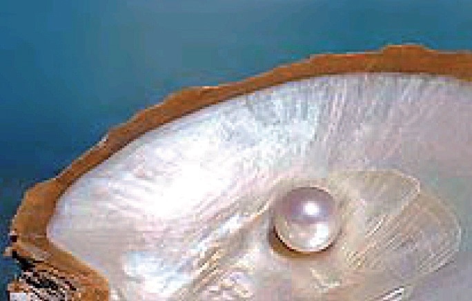
While pearls are generally known as high-fashion accessories, a wide selection of product with varying prices are available in the market. The cost of pearls is heavily dependent on their composition and provenance.
Pearls could be classified as natural, culture, or imitation. Natural pearls form when a small irritant is trapped inside a mollusk. The mollusk senses the irritant and starts depositing calcium carbonate minerals around the foreign object. As time passes, these minerals grown into pearls. Cultured pearls are formed in mollusks when seeding through the use of irritants is facilitated by humans; basically, pearls are cultured when human intervention is involved in the process. Meanwhile, synthetic pearls may be in the form of plastic or glass imitations as well as reconstructed calcium carbonate with a color coating.
The internal structure of pearls is dependent on their origin. For instance, natural seawater pearls are usually formed without a nucleus; meanwhile, cultured seawater pearls would often have a nucleus and a nacre layer that is 0.5 to 2 mm thick. Freshwater pearls, on the other hand, may or may have nuclei, but the most common variety available in the market would have none. Freshwater pearls could be distinguished from seawater pearls through their surface characteristics such as luster or patterning. Imitation pearls are composed of plastic or glass, making characterization easier since they lack calcium content.
X-Ray Analysis of Pearls
The optimized beam collimation provided by the mono-capillary x-ray guide tubes of the XGT-5000 instrument allows even large spherical samples such as pearls to be accurately analyzed, even enabling the parallel micro-beam to be unaffected by the significant curvature of the pearl’s surface. The elemental composition and internal structure of particles could be quickly and clearly visualized using the device. As such, the XGT is an ideal mechanism for the non-destructive investigation of pearls, particularly in identifying the possible presence of nuclei and conchiolin layers (via transmission imaging) in such and their characteristic composition (via XRF analysis).
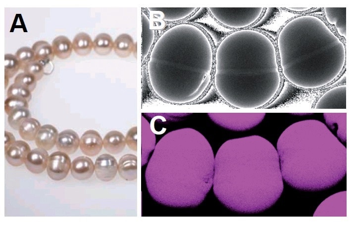
A) Chinese freshwater cultivated pearls, (B) transmission x-ray image and (C) calcium XRF image over a portion of the necklace
Because many imitation pears are made up of plastics, which are invisible to an XRF instrument, such could be distinguished easier from natural or cultured pearls that have calcium content.
Using the device, it is also possible to gain insight into the complex colorants used for these imitations. It was found that in some pearls, a strong presence of bismuth is evident. This is not surprising as there are a number of bismuth species used in typical pearlescent colorants.
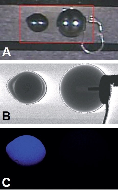
A) Optical image of pearls, cultured (left) and imitation (right), with mapping region shown in red, (B) transmission x-ray image, and (C) calcium XRF image.
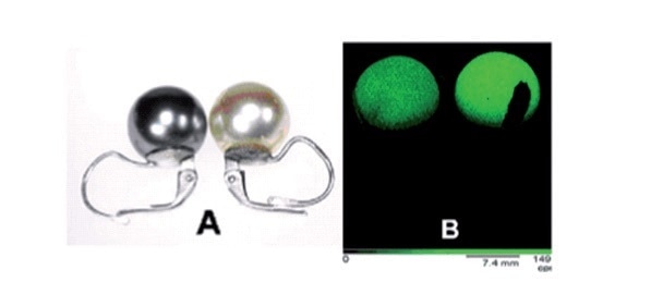
A) optical image of two imitation pearls, (B) bismuth XRF image
An analysis of the x-ray transmission images of two cultured pearls shows distinct internal structures that characterize various types of pearls. In particular, the clearly visible ring structure in the Tahiti seawater pearl is caused by a thick membrane of conchiolin. Meanwhile, Chinese freshwater pearls are cultured around a minute fragment of mollusk organic tissue. This is proven by a result showing no nuclei or conchiolin (nacre) layers in such type.
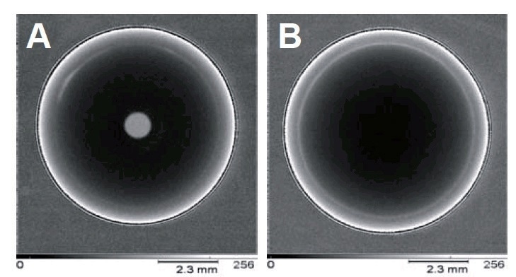
Transmission x-ray images of two cultured pearls, (A) high quality freshwater pearl from China, and (B) seawater pearl from Tahiti.
Conclusions
The fast mapping capability of the XGT-5000, with simultaneous XRF and transmission detection features, has enabled a range of pearl samples to be rendered analyzable. The relevant elemental and structural information acquired from natural, cultivated, and imitation pearls provide the researcher with useful information on the material’s composition and provenance. The optimized x-ray beam collimation provided by the HORIBA x-ray guide tube technology enables large pearls (with diameters of 1-2 cm) to be accurately probed for their internal structure and characteristics.

This information has been sourced, reviewed and adapted from materials provided by HORIBA.
For more information on this source, please visit HORIBA.