It is crucial to know what controls solid-state reactions, crystallization, phase stability, and solubility since the physical state can affect the pharmaceutical behavior of drug substances.
There are a number of techniques which have been utilized to measure the solid-state composition of pharmaceuticals, these include:
- Optical microscopy
- X-ray diffraction
- NMR
- Particle size analysis
- Thermal analysis
- Infrared (IR) spectroscopy
- Dissolution testing
In this industry, Raman spectroscopy is a newcomer as a very powerful characterization method. Indeed, Raman spectroscopy can supply qualitative and quantitative information of the polymorphy, with 1 µm spatial resolution when required.
The new generation in Raman technology supplies many advantages over the other methods. Samples can even be studied in transparent glass or plastic containers, as a non-destructive analysis.
Microscopic samples which are as small as 1 µm can be easily characterized, and little or no sample preparation is needed. Furthermore, polymorphic and pseudo-polymorphic phases in microscopic samples can also be mapped.
As the pelletizing can create pressure-induced polymorphic transformation this last point is crucial. In this article, the different polymorphic phases of carbamazepine, firstly on pure powder, and next after tableting, are discussed.
Instruments and Techniques
The HORIBA LabRAM SoleilTM is a Raman microscope providing high throughput with no compromise on resolution. This is due to the unique optical design of this microscope based on dielectric mirrors, with very low signal loss, coupled with high-quality gratings.
In order to acquire the best quality spectra necessary for polymorphy analyses, these outstanding characteristics are needed. By definition, the differences between the two polymorphic phases in the crystal modes, which can be characterized on the low Raman frequencies region.
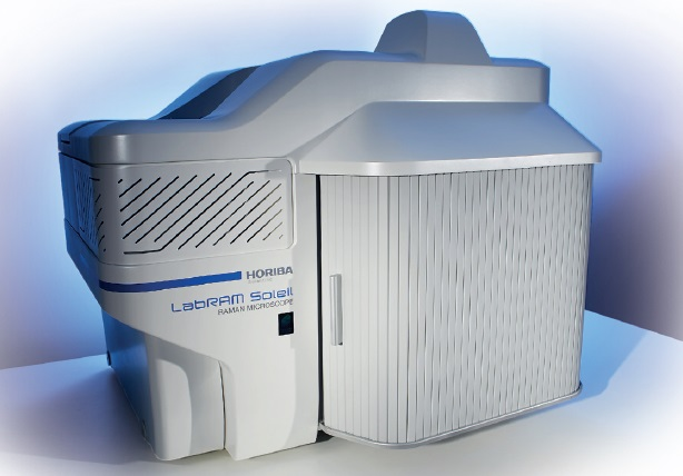
Figure 1. HORIBA LabRAM SoleilTM Raman microscope.
This means that difficulty for the discrimination of the phases is increased. It becomes easy to reach 30 cm-1 frequency and to characterize polymorphisms without additional options, because of the standard super low-frequency standard module available on LabRAM SoleilTM.
By automatically changing the angle of the injection/rejection edge filter this spectral range is achievable. Consequently, the Raman throughput is much better than through “classical” ultra low-frequency filters.
Results
Initially, different single spectra obtained on raw carbamazepine powder were compared and two different spectra are seen. These spectra are similar, as seen in Figure 2, except in the low-frequency range, below 50 cm-1.
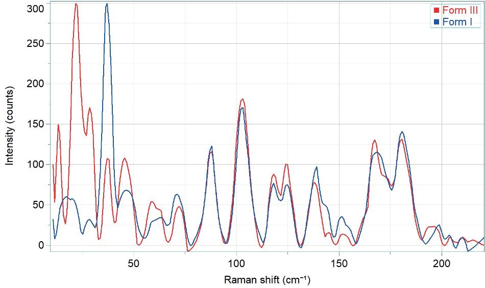
Figure 2. Low-frequency Raman spectra of Carbamazepine grains of powder.
In this specific range, on Form I a specific band of this crystal phase at 40 cm-1 is observed. This is exactly the type of band that helps to discriminate between polymorphisms. On raw products, Raman is an excellent tool to discriminate between two polymorphic forms.
Still, Raman microscopy can also be utilized to discriminate polymorphic forms on tablets or pellets. A homemade tablet of different forms of carbamazepine mixed with excipients was analyzed using a second method.
A Raman map was first utilized to identify carbamazepine particles in the tablet, and second, the polymorphic phases of these particles. It became easy to distinguish between the different forms I and III based on the spectral range 30-50 cm-1. Figure 3 shows the results on the polymorph distribution.
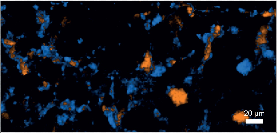
Figure 3. Carbamazepine distribution in tablet (blue: Form I, orange: Form III, black: excipients).
Conclusion
For polymorphic phases characterization of API, Raman microscopy is the ideal tool. The extremely low-frequency module which is available on HORIBA LabRAM SoleilTM as standard is perfect for acquiring crystal phase bands with no compromise on the acquisition time, which is a must-have for genuine product characterization.
Acknowledgments
Produced from materials originally authored by Thibault Brulé, Céline Eypert from HORIBA FRANCE SAS.

This information has been sourced, reviewed and adapted from materials provided by HORIBA.
For more information on this source, please visit HORIBA.