In an effort to meet energy reduction commitments, lithium-containing compounds and alloys are essential to make progress.
Though there have been undeniably enormous strides in developing these materials, what is even more impressive is that such improvement has essentially been achieved without a reliable method to discover or measure the distribution of lithium at the microscale.
Given that energy dispersive X-ray spectroscopy (EDS) is largely ineffective for elements with an atomic number lower than 4, finding a suitable quantitative method to map lithium in bulk materials remains highly sought after for microanalysts.
Recently, however, quantification of Li in the SEM was demonstrated via the use of a composition by difference method based upon EDS and quantitative backscattered electron imaging (qBEI).1
In order to generate quantitatively spatially resolved elemental maps of a MgLiAl alloy, Gatan chose to use the composition by difference method.
Mapping lithium in the scanning electron microscope (SEM) was possible using of Gatan’s OnPoint™ detector and EDAX’s Octane Elite Super EDS system.
Materials and Methods
Elements Z = 4 – 94 are quantified by the use of EDS in the composition by difference method, whilst qBEI defines the mean atomic mass (the qBEI signal being a function of atomic number for Z = 1 – 94). The difference in the two data sets is then used to calculate the fraction of ‘missing’ light elements (Z = 1 – 3).
The Ilion® broad beam argon ion polisher was used to prepare a MgLiAl, which was then analyzed using the OnPoint backscattered electron (BSE) detector and Octane Elite Super EDS system in a field emission SEM.
DigitalMicrograph® software was used to quantify the BSE data and to perform the calculation of aluminum, magnesium and, for the first time, lithium elemental maps.
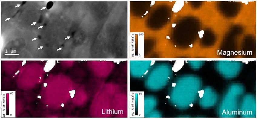
Figure 1. Secondary electron image and elemental metal fraction maps (by wt. %) of the same region of the MgLiAl alloy; white pixels are regions excluded from the analysis due to influence of topography (identified by arrows in the secondary electron image) shown here for clarity. Image Credit: Gatan Inc.
Summary
Quantitative mapping of single-digit mass percentages of Li took place in the SEM over a field of view of tens of microns with sub-micron spatial resolution. The insensitivity to the chemical bonding state and the lower detection limit makes this a very appealing technique for spatially resolved analysis of low atomic number elements.
References
1) J.A. Österreicher et al., Scripta Materialia 194 (2021) 113664

This information has been sourced, reviewed and adapted from materials provided by Gatan Inc.
For more information on this source, please visit Gatan Inc.