The revolutionary design of the Monarc™ system allows the spatial and spectral (color) information offered via the cathodoluminescence (CL) signal to be effectively and rapidly captured. These capabilities provide new and expanded insight into mineralogical processes.
Context
CL microscopy analyzes the light emitted by a gemstone or mineral when this is excited using an electron source. This technique is widely recognized as a practical and essential part of the microanalysis toolkit.
CL microscopy offers in-depth insight into an area’s geological history. This technique is, therefore, seeing wide-ranging utilization in the determination of mineral provenance, including geochronology and metamorphic alteration studies.
More recently, CL microscopy has also been employed in thermobaromtery as part of petrographic applications.
A large number of CL detectors collect spatial information in the form of unfiltered (black and white) images, though color CL imaging has widely replaced unfiltered imaging in recent years due to its extended access to valuable and easy to interpret information.
Despite its benefits and popularity, color imaging only provides limited spectral information, inhibiting quantitate analysis, the identification of trace elements and limiting the ability to determine their valence and structural position.
Hyperspectral imaging (spectrum imaging) has long been known to offer many benefits due to its capacity to collect the full range of spatial and spectral information in a single data set, collating this as spectrum image or hyperspectral data cube.
The acceptance and implementation of CL spectrum imaging in geosciences have remained low, however, primarily due to its slow acquisition speeds.
Materials and Methods
A typical scanning electron microscope (SEM) works by scanning the electron beam across a sample, collecting wavelength-resolved spectra (spectrum images) on a point-by-point basis.
The resulting spectrum images often exceeded a specific application’s spectral resolution requirements, but these tended to have limited spatial resolution due to the extended acquisition times.
The Monarc detector offers a novel acquisition standard, employing its ultra-fast detectors to collect a series of (aligned) wavelength-filtered maps which are in turn used to build the hyperspectral data cube. This method is able to significantly reduce acquisition times while continuing to enable high-spatial sampling.
Figure 1 features a comparison between two hyperspectral images of a polished zircon grain. These have been captured with a Monarc detector’s wavelength-filtered spectrum imaging and traditional wavelength-resolved modes.
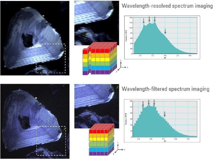
Figure 1. True-color representations of spectrum images captured by wavelength-resolved and wavelength-filtered modes of the Monarc detector. Both data sets were captured in 150 s. Image Credit: Gatan Inc.
A comparison of the extracted spectra reveals that it is possible to detect all spectral features, even in spite of the limited wavelength sampling in the wavelength-filtered approach (42 channels).
Monarc’s unique detection method is able to facilitate >70x higher spatial sampling, making it possible to investigate the sample’s fine banding structure.
Summary
The Monarc detector’s novel wavelength-filtered spectrum imaging mode allows the collection of hyperspectral data up to 100x faster - or with 100x higher spatial sampling - than other CL detectors.
This major advancement is anticipated to result in hyperspectral imaging superseding the traditional color and black and white CL imaging modes, empowering scientists to gain insight into even mineralogical processes via CL microscopy.

This information has been sourced, reviewed and adapted from materials provided by Gatan Inc.
For more information on this source, please visit Gatan Inc.