The CMS196 instrument, designed in association with Professor Bram Koster’s team from Leiden University Medical Centre (LUMC) in the Netherlands, is the outcome of four years of research and development.
The correlative stage was developed out of the necessity for a tool that uses fluorescence microscopy to help localise structures of interest in samples for high-resolution cryo-electron microscopy imaging. Over the years the system has evolved into an effective and efficient instrument that enables the imaging of cryo electron microscope images after light microscopy characterisation routinely.
A.J. Koster, Professor, Leiden University Medical Centre
CMS196V³ Cryo-CLEM Stage: Overview
The cryo-CLEM instrument—CMS196V³—allows a complete workflow of CLEM. Not only does it keep the sample vitrified through liquid nitrogen cooling but it also has established capabilities to safely transfer and handle cryo specimens and image them through optical microscopy. At the same time, it makes sure that the samples are free of contamination at any given time.
The built-in motorized, encoded stage of the new model, along with the latest LINK software helps create high resolution maps of the grid easily and rapidly. This allows a simple technique of coordinate mapping needed to identify the same specimen both in the fluorescence microscope and the EM.
While the CMS196V³ instrument was particularly developed to resolve the concerns relating to vitrification of EM grids from the fluorescent microscope into the cryo TEM without contamination and devitrification through condensation, it also had to be improved optically to facilitate the use of high NA lenses.
Up to three grids are placed into a uniquely developed cassette, which, in turn, is shifted from a plunge freezer into a compact sealed container. This container is subsequently placed into a pre-cooled dry sample loading chamber fixed on an upright fluorescent microscope. Then, using unique manipulation tools, the cassette is easily placed onto the viewing bridge.
Features/Highlights of the CMS196V³
- Automatic top-up liquid nitrogen is available to top up the chamber
- A self-contained cryo sample observation system keeps the specimen vitrified at a constant temperature of −196°C and prevents contamination
- Built-in motorized XY stage with position readout of better than 1 µm and optical encoders for high precision movements
- Startup time takes only 10 minutes, no mechanical connection between Dewar and sample mount, excellent stability, good long-term stability and low drift in the nanometer range
- A mounting option is available for a majority of research-grade, upright microscopes from all leading manufacturers
- XY mapping is fully automated
- Support for Auto-Grids (FEI), EM-grids, Bessy grids, Membrane carriers (Leica HPM100), CryoCapsule from CryoCapCell, planchettes and customized sample holders; there is no need to manage bare grids
- A self-aligning magnetic sample cassette system is available for up to three sample grids
- Can be linked to high pressure freezing and plunge-freezing sample preparation procedures
- Sample cassette holder enables safe and contamination-free loading, transfer and storage of samples
- Built-in condenser optics enables phase contrast and transmitted light bright-field
- Built-in sensors help monitor the temperature of samples
- Full day of individual operation with a 3-L autofill LN2 Dewar provided as an option
- Fluorescence observation with up to 0.9 NA
- Automated heating “bake-out” cycle removes moisture
- LCD display with button control, full software control through a USB interface and standalone joystick operation
- Over 50 systems have been proven in the field across the world, making Linkam Scientific the market leader in the domain of Cryo-CLEM
Correlative microscopy - automated mapping of an EM grid at high resolution
CMS196M automated mapping of an EM grid at high resolution. Video Credit: Linkam Scientific Instruments
Why Cryo-CLEM?
While electron microscopy (EM) offers structural data at an excellent resolution, it can provide only a limited understanding of chemical and biological processes because of the limitations in sample preparation and staining procedures. By contrast, fluorescence microscopy is a highly sensitive technique that helps detect genetic, chemical and biological events and processes within living cells.
Now, Cryo-CLEM brings it all together: it is a novel, emerging method that integrates the individual benefits from both EM and fluorescence by imaging the location of the same specimen using both methods and superimposing the complementary data.
We are using the cryo-correlative stage to screen cells trapped in a thin layer of ice prior to imaging the samples at synchrotrons in Oxford, Barcelona and Berlin. The team at Linkam Scientific have helped to drive forward our research, quickly adapting and refining the technology to deliver an elegant, flexible, user-friendly stage.
Dr Lucy Collinson, Head of Electron Microscopy, The Francis Crick Institute
Imaging of Vitrified Samples Under Cryo-Conditions
To make sure that a biological specimen is compatible with the vacuum conditions present in EM and that the structural detail is preserved, specimens are integrated into vitrified “glass-like” ice and these should be maintained below −140°C.
Any contact with atmospheric moisture should be prevented because ice crystals would form quickly and thus contaminate the specimen. Under cryo-conditions, the structural detail of the fluorescence signals is preserved and photobleaching is considerably decreased.
Linkam Co-Ordinate Transform Tool
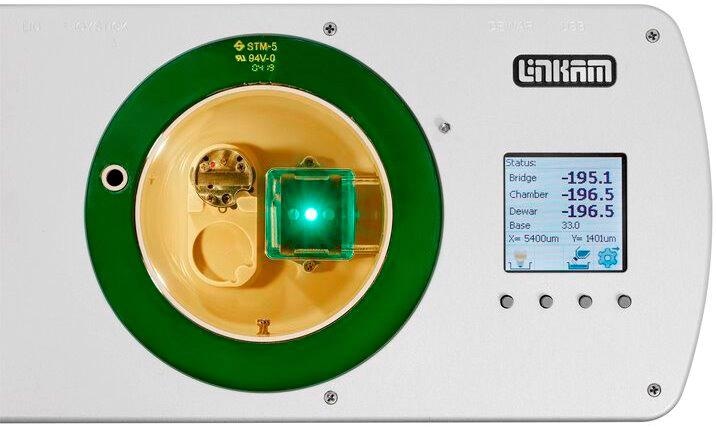
Image Credit: Linkam Scientific Instruments