As fascinating as microscopic images are, ZEN Intellesis’ true worth lies in the information they give. Image segmentation is one of the most difficult problems in microscopy, and it serves as the foundation for all future image analysis procedures.
Even non-experts can quickly develop solid and repeatable segmentation results with ZEN Intellesis’ Python-based machine-learning algorithms, including pixel categorization and deep learning. Users may now configure the software once, and ZEN Intellesis will automatically segment a batch of hundreds of photos. Users can save time while reducing user bias.
Highlights
Use Deep Learning to Segment Images
Users often need to segment distinct types of items in several photos to acquire trustworthy data from them. With ZEN Intellesis, users can now prepare the software on a few photos using personal knowledge.
The advanced machine learning algorithms (including Deep Learning) of the software module ZEN Intellesis will then perform all of the time-consuming segmentation procedures on the hundreds of comparable photos.
Even complex multidimensional, multi-modal data may be studied with the Python-powered tools, which include pixel categorization with real multichannel extracting features and segmentation using pre-trained networks. Users may also integrate and use their own deep learning models with the software.
Enjoy Smooth Workflow Integration
Machine learning is made simple using the software module ZEN Intellesis: Simply install the image, create the classes, label pixels, prepare the model and segment the image. Users can even incorporate a pre-trained model for typical tasks like Nucleus Segmentation, which can be used to segment and analyze complete datasets.
Additionally, users can train their own network and import it into ZEN (a commercial service for creating customized pre-trained networks is also available).
Intellesis is scriptable and is completely integrated into the Image Analysis Framework in ZEN Blue and ZEN core Imaging Software. This integration ensures that all of the important Metadata remains connected and accessible for subsequent processing processes.
Analyze Multi-Modal Images from Many Sources, in Many Formats
With the ZEN Intellesis software module, users can effortlessly segment multidimensional images from a wide range of imaging sources, including:
- Label-Free Microscopy
- Confocal Microscopy
- X-Ray Microscopy
- Electron Microscopy
- Fluorescence Microscopy
- Super Resolution Microscopy
- Widefield Microscopy
- Light Sheet Microscopy
With ZEN Connect, users may merge images from several microscopes on the same material and use the outcomes with ZEN Intellesis to extract information that is even more useful. To segment the structures of interest, the software will employ feature representations from all modalities at the same time. ZEN Intellesis can segment all Bio-Formats compatible images by simply importing OME-TIFF or TXM images or by using the third-party import function.
Application Example Life Sciences
1. Scratch Assay
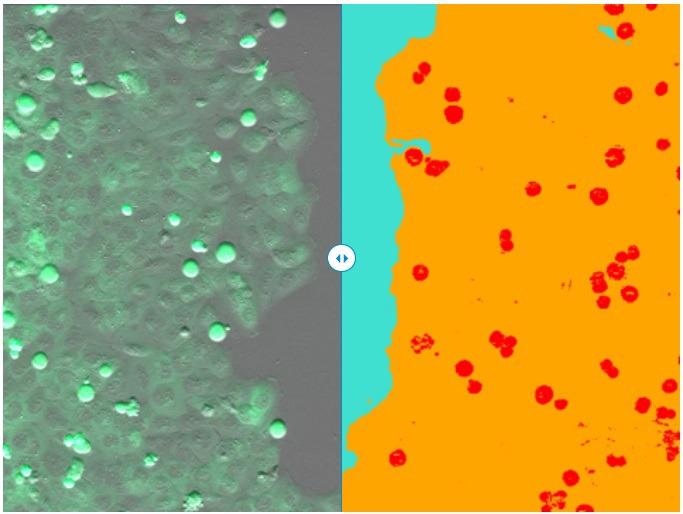
(left) Single frame of recorded time-lapse movie. (right) Segmentation result of ZEN Intellesis - scratch area (turquoise), cell layer (orange), and mitotic cells (red). Image Credit: Carl Zeiss Microscopy GmbH
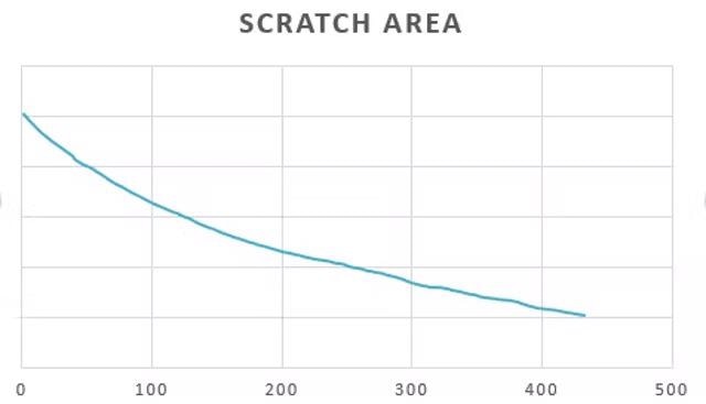
Based on ZEN Intellesis segmentation results, the size of the scratch area was measured over time using the ZEN Image Analysis module. Image Credit: Carl Zeiss Microscopy GmbH
2. Spines and Dendrites
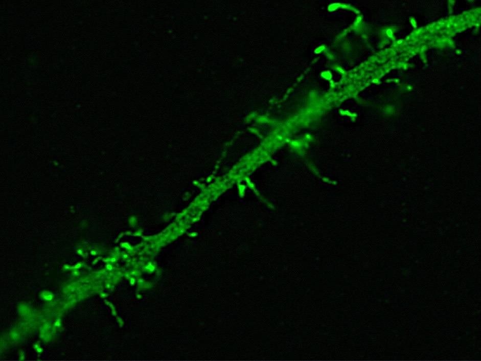
Dendrite of a neuron expressing Green Fluorescent Protein. Image acquired with structured illumination on an Elyra PS.1 showing spines on a dendrite. Image Credit: Carl Zeiss Microscopy GmbH
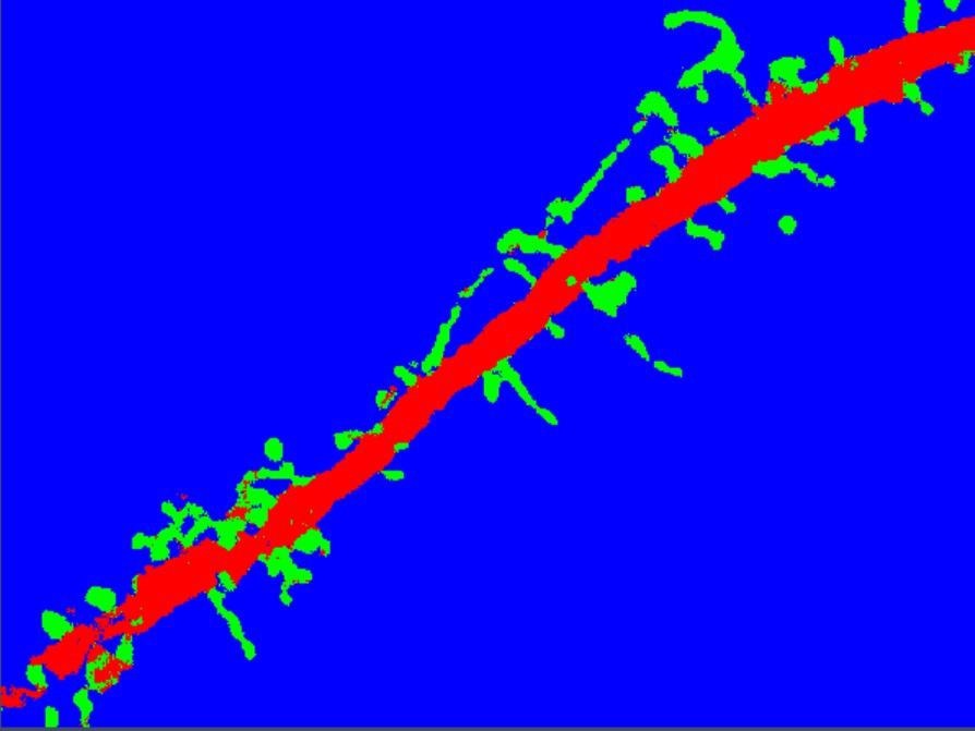
Segmentation with ZEN Intellesis results in a clear separation of spines (green) from dendrite (red) and background (blue). Image Credit: Carl Zeiss Microscopy GmbH
3. Drosophila
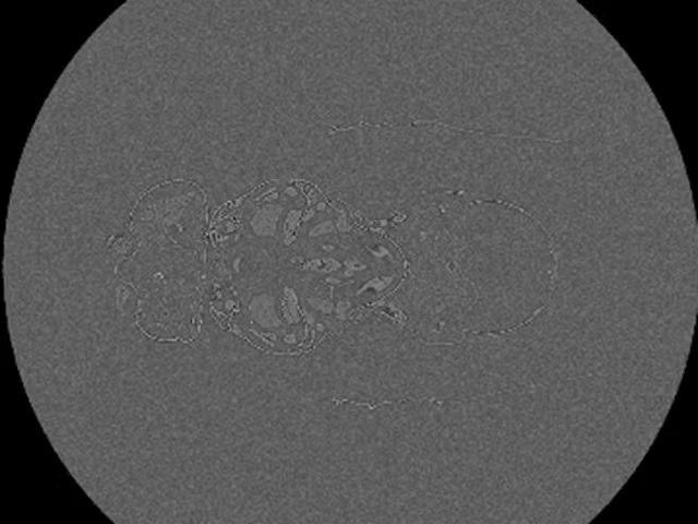
X-Ray micrograph of a Drosophila melanogaster. 1400 slide z-stack of the whole fruit fly acquired with ZEISS 520 Versa. Image Credit: Carl Zeiss Microscopy GmbH
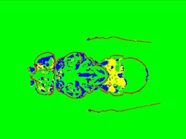
Segmentation result obtained with ZEN Intellesis – exoskeleton (red), inner structures (blue and yellow), background (green). Image Credit: Carl Zeiss Microscopy GmbH
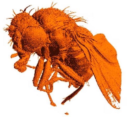
Rendered 3D model based on exoskeleton class. Image Credit: Carl Zeiss Microscopy GmbH
Application Example Materials Analysis
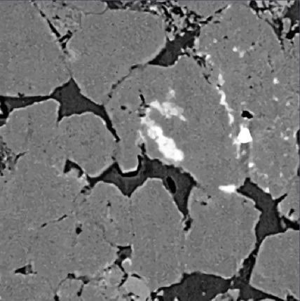
XRM Sandstone – Original. Image Credit: Carl Zeiss Microscopy GmbH
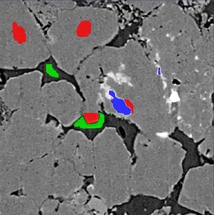
XRM Sandstone – Labeled. Image Credit: Carl Zeiss Microscopy GmbH
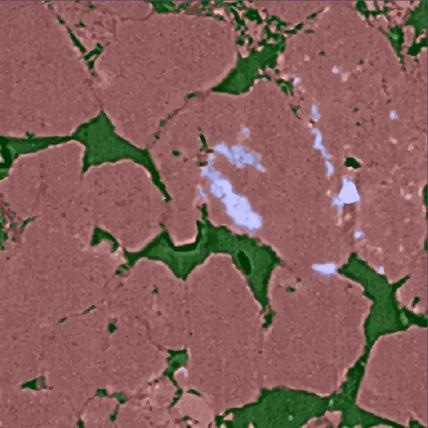
XRM Sandstone – Trained & Segmented. Image Credit: Carl Zeiss Microscopy GmbH
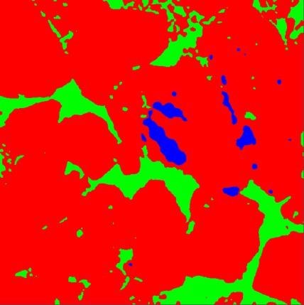
XRM Sandstone – IP Function Output. Image Credit: Carl Zeiss Microscopy GmbH
How to Segment the Images
See it With Own Eyes: Download the Free Trial Version
The images contain the truth: Download the free 30-day trial edition of ZEN Intellesis by registering today. Users will be able to use the entire capabilities of this software module for deep learning picture segmentation:
- Train the software
- Label the classes in the specimen
- Define the classes in the sample
- Load a large variety of image files
- Segment images