The inLux™ SEM Raman interface equips a variety of SEM chambers with high-quality Raman functionality. Now, users can gather Raman spectra and simultaneously image inside their SEM.
Users can rest assured that they are receiving accurate co-location when comparing Raman images and SEM images since the sample remains static during the Raman data collecting and SEM imaging processes.
A wide range of Raman capabilities are available through the inLux interface. Users can generate 2D and 3D confocal Raman images or collect spectra from single or multiple points. This data can be overlaid on the SEM image to give the context of chemical information to the microscope image.
Users of the inLux interface can analyze volumes larger than 0.5 mm in each axis and it features a fully encoded position control that ensures accurate Raman imaging down to 50 nm.
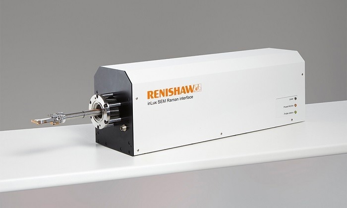
Image Credit: Renishaw plc-Spectroscopy
Key Benefits
- Non-invasive: At the click of a button, the inLux probe can be fully retracted, ensuring that the probe does not interfere with other SEM functions or workflows when it is not being used
- Sample viewing: Using large-area optical imaging and montaging, the sample can be observed and areas of interest can be targeted
- Automated: The Raman analysis of challenging samples can be achieved via one-click switching of the laser wavelengths
- Information-rich: Raman, photoluminescence (PL) and spectral cathodoluminescence (CL) analysis is performed simultaneously and co-located with SEM imaging
- Configurable: Additional options include a CL module and up to two distinct excitation laser wavelengths
- Universal: Without requiring any SEM modification, the inLux interface can be mounted on a wide variety of SEMs from various manufacturers with various chamber diameters
- Determine distribution: It is simple to quantify sample heterogeneity as confocal Raman images can be produced as a standard
Video Credit: Renishaw plc-Spectroscopy
inLux Interface Applications
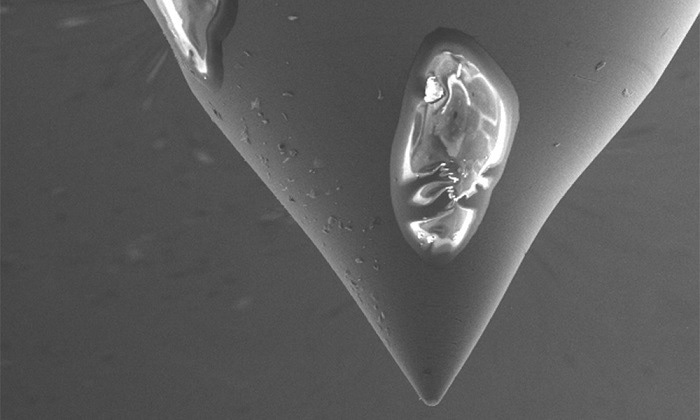
Image Credit: Renishaw plc-Spectroscopy
Identifying Contaminants
As a non-contact and non-destructive technique, Raman spectroscopy can provide highly specific chemical information, which means that it is the ideal method for identifying contaminants. Raman spectroscopy is especially effective for analyzing organic and carbon contaminants that are challenging to distinguish using elemental analysis.
A SEM can be used to locate and study the morphology of small contaminant particles that cannot typically be resolved via optical microscopy. Next, without having to change the sample, these particles can be specifically targeted for Raman analysis utilizing the inLux interface to provide chemical analysis.
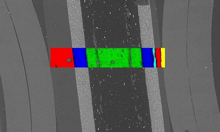
Image Credit: Renishaw plc-Spectroscopy
Materials Analysis
The size, shape, or thickness of a material can influence a lot of its new features. Graphene, nanorods and nanotubes are examples of where the high magnification of scanning electron microscopy is vital when it comes to the visualization of the sample.
Raman analysis can reveal the physical qualities of the material in addition to its chemical and structural composition. The inLux interface is able to produce Raman images that have crystallinity, strain, and electronic properties that can be correlated to those from the SEM.
Connect to Different Renishaw Raman Systems
The research-grade Raman spectrometers and software from Renishaw are used in conjunction with the inLux interface. This combination ensures thorough processing and analysis capabilities and is incredibly user-friendly.
The inLux interface can assist users in making the most of the SEM for a variety of uses, including identifying industrial contamination and academic research.
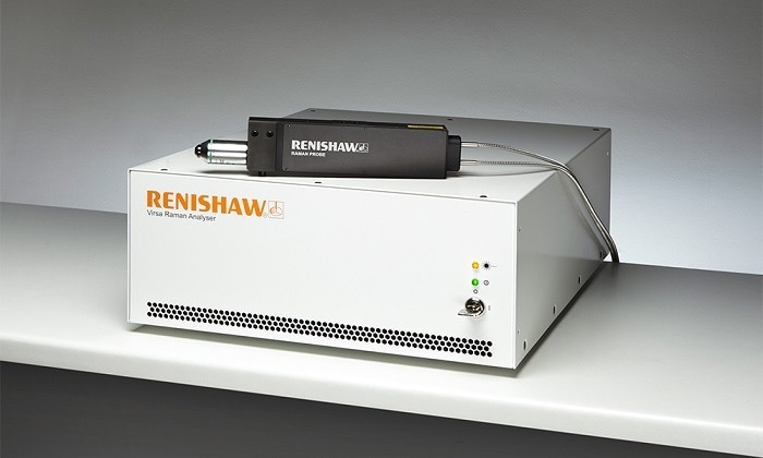
Image Credit: Renishaw plc-Spectroscopy
Virsa™ Raman Analyzer
The Virsa Raman analyzer can be connected to the inLux interface for specialized Raman analysis. The Virsa analyzer is considered a cost-effective, compact solution to in-SEM Raman analysis without compromising on the high sensitivity and spectral resolution that is expected of a research-grade Raman system in a rack-mounted body.
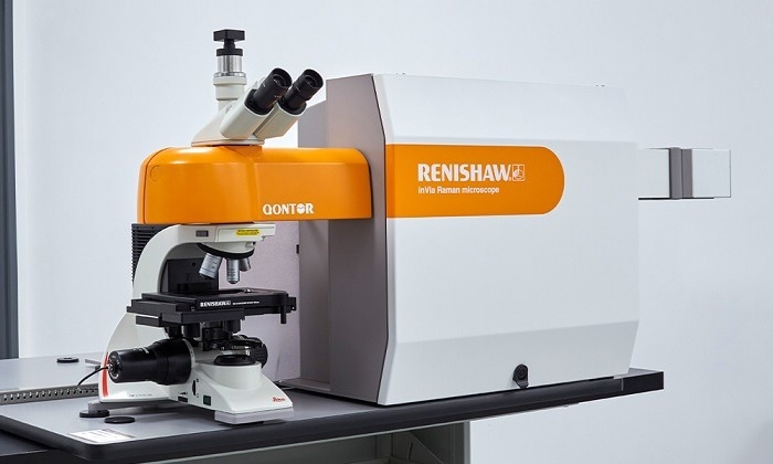
Image Credit: Renishaw plc-Spectroscopy
inVia™ Confocal Raman Microscope
The world’s most popular research-grade Raman microscope can now include in-SEM analysis by connecting the inLux interface to the inVia confocal Raman microscope. The inVia microscope ensures world-leading performance and sensitivity in a configurable range of laser excitation wavelengths, detectors and gratings.
Any Raman active material can be analyzed using this device. The inVia microscope can be used independently for Raman analysis. When in situ measurements are not necessary, the SEM can subsequently be made available to other users.
Specifications
Source: Renishaw plc-Spectroscopy
| Parameters |
Value |
| Mass |
< 20 kg |
| Fiber optic cable length |
4.6 m |
| Compatible Raman spectrometers |
Renishaw inVia confocal Raman microscope, Renishaw Virsa analyzer |
| Compatible SEM models |
Compatible with models from all major SEM suppliers |
| SEM port requirement |
Requires free SEM side or rear port |
| SEM performance |
The inLux interface does not require any SEM modification and can be fully retracted when not in use, thus it does not interfere with SEM performance or that of other accessories |
| Movement control |
Trackpad, WiRE software |
| Contact protection |
Touch sensor, safe working volume monitored using absolute encoders |
| Laser safety |
Laser interlocked to chamber vacuum |
| Raman mapping/imaging |
Supplied as standard |
| Fiber optic module selection |
Up to two different laser excitation wavelengths + optional cathodoluminescence module |
| Available laser excitation wavelengths |
405 nm, 532 nm, 660 nm, 785 nm (others available on request) |
| Laser switching |
Automated, motorized, and software controlled |
| Lateral spatial resolution |
< 1 µm @ 532 nm |
| Confocal performance |
< 6 µm @ 532 nm |
| Spectral resolution |
See the spectrometer specification sheet |
| Dimensions |
W 804 mm x H 257 mm x D 215 mm |