Understanding the mechanisms by which the brain functions is one of the most complex challenges in science. One important aspect is the electrical conduction of stimuli in nerve cells. In order to study neuronal circuits, a sharp metal electrode is usually inserted into the brain to introduce a current. However, the response does not reflect the highly complex activation patterns of natural nerve stimuli. In addition, the direct current applied in this fashion causes damage to tissue through undesired electrochemical side reactions.
Collaboration between neuroscientists and nanomaterials researchers at Case Western Reserve University (Cleveland, Ohio, USA) has resulted in the development of a technique that is both gentler and elicits more natural nerve impulses. As reported in the journal Angewandte Chemie, the technique is based on a micropipette coated with semiconductor nanoparticles that activates neurons in brain tissue with visible or infrared (IR) light. In contrast to conventional electrodes, these photoelectrodes require neither wires nor electrical power.
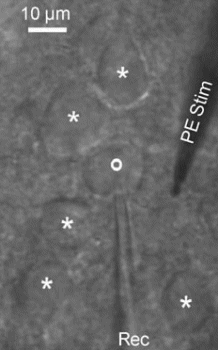
The team led by Ben W. Strowbridge and Clemens Burda coated the interiors of extremely finely drawn-out glass micropipettes with lead selenide nanoparticles. Lead selenide is a semiconductor that is activated by IR light. As in solar cells, irradiation “catapults” firmly bound electrons out of the valence band and into the conduction band of the semiconductor, where they can move freely. This leads to charge separation and thus to an electrical potential. With a suitable laser, defined processes elicited by short light pulses set off corresponding electrical pulses in the micropipette. An electrical field is thus formed around the pipette, which can then be used by the researchers to stimulate neurons in rat brain samples with a high degree of time-resolution. Measuring electrodes could then be used to record the natural activation patterns of very similar nerve impulses.
Samples of the olfactory bulb (a region of the brain involved in processing smell) and the hippocampus (part of the cerebrum important in the transfer of contents from short-term to the long-term memory) were examined. Neither toxic effects nor damage to the nerve cells were observed after repeated stimulation.
By using these new photoelectrodes, the cooperation of nerve cells can be studied. However, therapeutic applications are also possible: the probes could be used to activate individual regions of the brain or damaged or cut nerves to restore function – without the need for disturbing wires.