Feb 17 2014
A team of EPFL researchers have developed a mass spectrometry method that allows two-dimensional imaging of molecules on a surface.
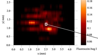 A "heatmap" generated with ESTASI/MSI ©2014 EPFL/LEPA
Mass spectrometry imaging (MSI) is a method that allows the localization of biological molecules or drugs in tissue. However, the most commonly used techniques require chemical preparation and have to be performed in vacuum, costing time and money. Publishing in Analytical Chemistry, a team of EPFL scientists have developed a MSI method that can be carried out efficiently in normal conditions, and can be applied to a wide range of molecules.
Mass spectrometry is one of the most powerful tools in analytical chemistry. Although it exists in many forms, the principal is essentially the same: Identifying unknown molecules based on their mass. Mass spectrometry is usually preceded by the development of ionization techniques. For biological molecules Common ionization methods include matrix-assisted laser desorption ionization and electrospray ionization. Both methods produce intact molecular ions to be separated and detected by a mass spectrometer, which then analyzes them according to their mass and identifies what the original sample is made of.
A team of researchers at EPFL’s Laboratory of Physical and Analytical Electrochemistry (LEPA) has developed a new ambient ionization technique called electrostatic spray ionization (ESTASI), and used it with mass spectrometry to gather chemical information across a two-dimensional surface. In this approach, the whole sample is placed on a platform and a wetting capillary delivers an acidic solution to the sample area to extract the molecules of interest for ESTASI. The wetting capillary acts as a scanning probe across the sample area, and the data are collated with a computer that generates a kind of “heat map” of the sample, with the distribution of localized chemical information overlain on it.
Using this technique, the scientists were able to analyze organic molecules, peptides, proteins, and even cells. The imaging resolution is determined by the inner and outer diameter of the wetting capillary, but under optimal conditions it could be better than 110 μm. The method itself requires equipment that can be portable, which also makes it easily accessible to mass spectrometry labs around the world.
The EPFL researchers are currently testing their novel method on the imaging of biological tissues. However, given its success, they expect that it will find broad application in chemical and biochemical analysis, and that it can be further expanded and improved with additional development and optimization.
Reference
Qiao L, Tobolkina E, Lesch A, Bondarenko A, Zhong X, Liu B, Pick H, Vogel H, Girault HH. 2014. Electrostatic Spray Ionization Mass Spectrometry Imaging. Analytical Chemistry DOI: 10.1021/ac4031779