Posted in | News

Combining Raman Microscopy with Scanning Electron Microscopy (SEM) To Study Inorganic And Mineral Samples At The Geological Institute Of Romania, Bucharest.

The Geological Institute of Romania, located in Bucharest, was founded in 1906. It is famous for its museum which hosts a collection of more than 80,000 samples of rocks, fossils and minerals from all over Romania.

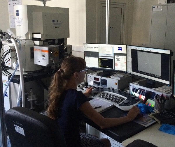
Oana-Claudia Barbu and Daniel Bîrgăoanu are both PhD students and research assistants in the electron microscopy laboratory, known as MICROCOSMOS, where they study inorganic and mineral samples. They use Renishaw’s structural and chemical analyser (SCA) interface to bring the Raman analysis capabilities of the inVia™ confocal Raman microscope to their scanning electron microscope (SEM). The inVia and SCA interface provide an in-SEM analytical technique that allows co-located chemical information and the flexibility of a fully functioning Raman microscope.

Ms Barbu commented, “This allows us to perform high resolution analysis on geological samples using energy dispersive spectroscopy (EDS) and Raman spectroscopy. Our Zeiss SEM is also equipped with electron back scattering (EBSD) and wavelength dispersive (WDS) detectors to give even better correlation of results. The system is easy to use and has user-friendly analysis software (WiRE 4™) that provides us with high definition chemical images from our very large Raman datasets. We greatly appreciate the strong support we have received from Renishaw.”

An example, typical of their work, is the study of mining waste to better understand its composition. In both the Raman spectrum and EDS analysis below, the metallic mineral content is identified as pyrite (iron sulphide, FeS2).

Please visit www.renishaw.com/earthsciences for further details on how Raman microscopy is being used in earth sciences applications. For more details of Renishaw’s co-located SEM-Raman system, visit www.renishaw.com/SEMRaman

Citations

Please use one of the following formats to cite this article in your essay, paper or report:

  • APA

    Renishaw plc - Spectroscopy. (2017, January 13). Combining Raman Microscopy with Scanning Electron Microscopy (SEM) To Study Inorganic And Mineral Samples At The Geological Institute Of Romania, Bucharest.. AZoM. Retrieved on April 26, 2024 from https://www.azom.com/news.aspx?newsID=47095.

  • MLA

    Renishaw plc - Spectroscopy. "Combining Raman Microscopy with Scanning Electron Microscopy (SEM) To Study Inorganic And Mineral Samples At The Geological Institute Of Romania, Bucharest.". AZoM. 26 April 2024. <https://www.azom.com/news.aspx?newsID=47095>.

  • Chicago

    Renishaw plc - Spectroscopy. "Combining Raman Microscopy with Scanning Electron Microscopy (SEM) To Study Inorganic And Mineral Samples At The Geological Institute Of Romania, Bucharest.". AZoM. https://www.azom.com/news.aspx?newsID=47095. (accessed April 26, 2024).

  • Harvard

    Renishaw plc - Spectroscopy. 2017. Combining Raman Microscopy with Scanning Electron Microscopy (SEM) To Study Inorganic And Mineral Samples At The Geological Institute Of Romania, Bucharest.. AZoM, viewed 26 April 2024, https://www.azom.com/news.aspx?newsID=47095.

Tell Us What You Think

Do you have a review, update or anything you would like to add to this news story?

Leave your feedback
Your comment type
Submit

While we only use edited and approved content for Azthena answers, it may on occasions provide incorrect responses. Please confirm any data provided with the related suppliers or authors. We do not provide medical advice, if you search for medical information you must always consult a medical professional before acting on any information provided.

Your questions, but not your email details will be shared with OpenAI and retained for 30 days in accordance with their privacy principles.

Please do not ask questions that use sensitive or confidential information.

Read the full Terms & Conditions.