In this study, COXEM Co., Ltd. analyzed cosmetic samples using Scanning Electron Microscopy (SEM) to investigate their surface morphology and compositional characteristics. The high-resolution imaging capabilities of SEM allowed for detailed observation of microstructures. These findings offer valuable insights into the physical and chemical properties of cosmetic materials, contributing to a better understanding of their performance and potential applications in product development.
This study explores the surface properties of cosmetics using COXEM’s new Tabletop Scanning Electron Microscope (SEM), the EM-40, providing insights into their microstructure and composition.

Cosmetics. Image Credit: COXEM Co. Ltd.
Introduction
The history of cosmetics dates back to ancient civilizations, with the Egyptians pioneering their use for skincare and beauty through natural ingredients. Subsequently, in Greek and Roman societies, cosmetics evolved as tools for aesthetic expression and markers of social status.
During the Middle Ages, the use of cosmetics was restricted for religious reasons, but they regained prominence during the Renaissance and modern periods, fueled by advancements in beauty practices and scientific knowledge. In contemporary times, industrialization and technological innovation have led to the diversification and widespread accessibility of cosmetics, establishing the industry as a vital component of the global market. With the rising popularity of gender-neutral cosmetics, the use of cosmetic products has become increasingly common across all ages and genders.
This study aims to analyze four essential cosmetic products frequently used in daily makeup routines through SEM. This investigation seeks to provide a deeper understanding of these widely utilized cosmetics' structural and compositional characteristics.
Materials and Sample Preparation
Four types of cosmetics - foundation, mascara, eyebrow pencil, and lipstick - were prepared for analysis. Each sample was placed on a sample holder and left to dry at room temperature for approximately 24 hours. Subsequently, the samples were coated using an ion sputter coater (SPT-20, COXEM., Ltd) with an Au target under conditions of 3 mA and 400 seconds. After completing the coating process, sample preparation was finalized, and observations were conducted using a scanning electron microscope (EM-40).
Cosmetics were prepared for surface analysis using COXEM’s Tabletop SEM, the EM-40. This SEM operates at a magnification of up to 250,000x and an accelerating voltage of up to 30 kV, enabling detailed examination of the cosmetic’s surface. The samples were analyzed to assess their microstructure and elemental composition, providing a comprehensive understanding of their properties.
Results
SEM images taken at 200x and 10,000x magnification have unveiled the complex microstructure of cosmetics. These images reveal the presence of skin and morphological features embedded in the surface of the cosmetics. The high-resolution imaging capabilities of COXEM's EM-40 SEM have also enabled a detailed analysis of the cosmetic`s surface, providing valuable insights that can enhance our understanding of its biology and potential applications in scientific studies.

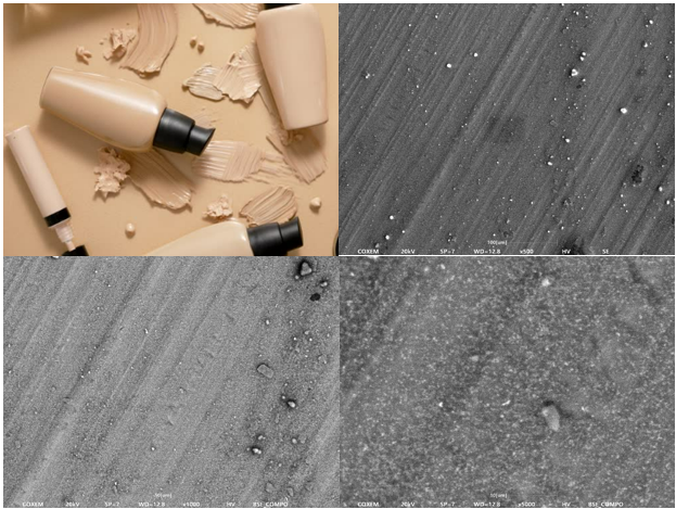
Foundation. Image Credit: COXEM Co. Ltd.
The SEM analysis of the foundation showed mostly small particles. The BSE images highlighted contrast differences, suggesting a mix of particles with different compositions. This indicates that, in addition to its main ingredients, the foundation likely includes extra components designed to enhance its UV-blocking properties. These UV-blocking agents appear to account for the variety in the foundation's composition.

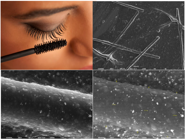
Mascara. Image Credit: COXEM Co. Ltd.
The SEM analysis of the mascara revealed the presence of clumped fibrous structures, presumed to be fiber components within the mascara formulation. Upon magnification at 5000x, small particles were observed on the surface of these fibrous structures, indicating a complex microstructure likely contributing to the mascara's performance characteristics.

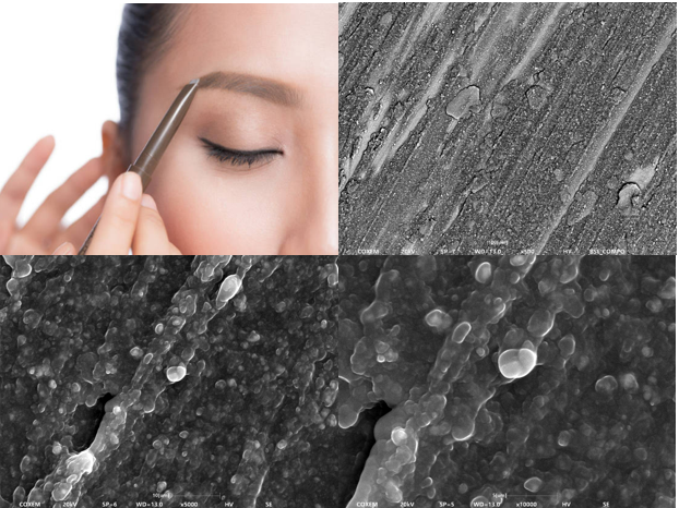
Eyebrow. Image Credit: COXEM Co. Ltd.
The SEM analysis of the eyebrow pencil revealed a pattern of small, spherical particles distributed across the surface upon application. The particles exhibited an irregularly clustered morphology while maintaining a uniform overall distribution, a characteristic that is particularly notable and may contribute to the product's application properties.

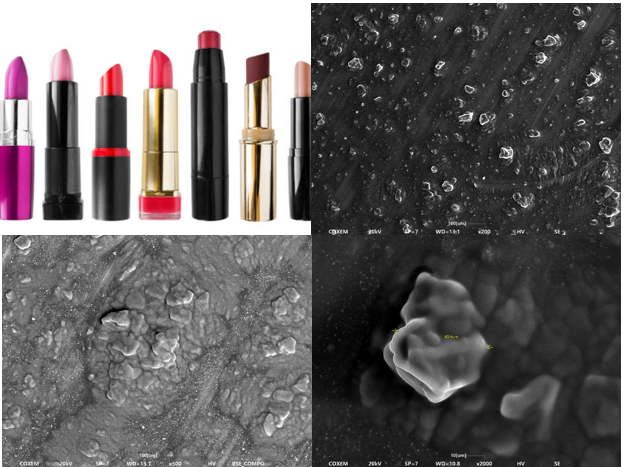
Lipstick. Image Credit: COXEM Co. Ltd.
SEM analysis of the lipstick revealed irregularly shaped particles within its characteristic smooth and soft texture when spread. These particles were observed to either cluster together or detach from the formulation, displaying a dispersed pattern. This distribution provides insights into the microstructural properties of the lipstick's formulation.
Discussion
The analysis has shown that the surface structure of cosmetics is distinguished by a uniform pattern. Leveraging COXEM's Tabletop SEM, the EM-40, researchers can capture high-resolution images of the cosmetic's surface, enabling detailed observation of its external structures at micro and nano scales. This advanced imaging capability is particularly useful for studying morphological changes, surface textures, and structural features that are not detectable with conventional optical microscopy. These insights contribute significantly to our understanding of cosmetic science and its applications in various research fields.
Conclusions
The SEM analysis of the selected cosmetics - foundation, mascara, eyebrow pencil, and lipstick - provided valuable insights into their microstructural characteristics.
The foundation showed small, compositionally diverse particles likely linked to its UV-blocking components. Mascara exhibited fibrous structures with surface particles, suggesting enhanced volumizing properties. The eyebrow pencil displayed uniformly distributed spherical particles, contributing to its smooth application. Lastly, the lipstick revealed irregular particles within a soft matrix, reflecting its unique texture.
COXEM’s Tabletop SEM, the EM-40, is an invaluable tool for examining cosmetics, offering high-resolution images that reveal intricate surface structures. This capability is essential for understanding the connection between a product's microstructure and its functional properties, supporting both physical and chemical research in cosmetics development.

This information has been sourced, reviewed and adapted from materials provided by COXEM Co. Ltd.
For more information on this source, please visit COXEM Co. Ltd.