Scanning Electron Microscopy (SEM) offers high-resolution chemical microanalysis and imaging at the micro- and nano-scale.
Image Credit: Hitachi High-Tech Europe
When combined with advanced cross-sectioning techniques such as ion-beam milling, electron microscopy allows researchers to examine structures, morphology, and material distribution precisely.
Furthermore, sophisticated image analysis methods can transform these images into quantitative data that can be directly correlated with battery performance characteristics.
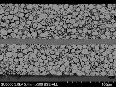
High-quality cross-section of LiB cathode (NMC) prepared with broad Argon ion milling. Image Credit: Hitachi High-Tech Europe
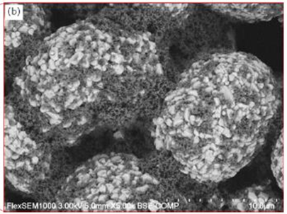
Studying binder distribution on cathode active material (CAM). Image Credit: Hitachi High-Tech Europe
Particle Morphology and Size
SEM provides insights into the size distribution and morphology of electrode particles. For instance, irregularly shaped particles, cracked particles, or agglomerates may impede ion diffusion and contribute to capacity fade.
Distribution of Active Materials
Electron microscopy can elucidate the distribution of active materials, such as lithium compounds or transition metal oxides, within the electrode matrix.
The distribution of other components, including polymeric binders and carbon nanotube-based conductive agents, can also be characterized.
Pore Structure
Imaging can reveal the internal pore structure of the electrode material, detailing the size, shape, and distribution of pores that are critical for electrolyte penetration and ion transport.
Interface Characteristics
The electrode-current collector interface and electrode-electrolyte interface (SEI) are vital for ion transport and electron conduction. Coatings, cracks, or other defects that may impact battery performance can be examined.
Structural Changes During or After Cycling
By comparing images taken before and after cycling, electron microscopy can clarify structural changes such as particle cracking, delamination, or the evolution of the electrode/electrolyte interface. Additionally, solid-state batteries can be studied in situ to observe lithium transport during charging and discharging cycles.

This information has been sourced, reviewed and adapted from materials provided by Hitachi High-Tech Europe.
For more information on this source, please visit Hitachi High-Tech Europe.