Image Credit: Tokyo Institute of Technology
Measuring brain activity can help with the diagnosis of epilepsy and other neuropsychiatric diseases. Electroencephalography (EEG) is the least invasive of the methods used. Electrodes are frequently put on the scalp during EEG recordings. However, the resolution of EEG is limited by the fact that electrical impulses from the brain are attenuated and distorted by the time they reach the scalp.
Electrocorticography (ECoG), on the other hand, includes the implantation of neural electrodes directly on the surface of the brain. ECoG electrodes give better recordings of brain activity because they are in direct touch with the region of interest.
Furthermore, electrical pulses can be sent via them to activate specific groups of neurons to manage epileptic episodes. However, typical ECoG electrodes have a significant disadvantage.
They frequently do not correspond to the mechanical characteristics and curvature of brain tissue, resulting in increased brain pressure and other negative consequences. Although flexible brain electrodes have been created to address this issue, they are either not durable or need sophisticated production methods.
To solve these issues, a Tokyo Institute of Technology (Tokyo Tech) research team led by Associate Professor Toshinori Fujie created a novel type of flexible neural electrode. Their design and findings, which were just published in Advanced Materials Technologies, have the potential to revolutionize ECoG recordings and direct neural stimulation.
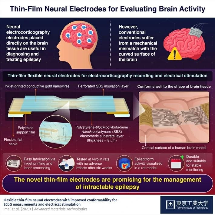
The suggested electrode’s substrate is a thin layer of a flexible material termed polystyrene-block-polybutadiene-block-polystyrene (SBS). The researchers created conductive wire on the electrode with gold nanoink using an inkjet printer. Finally, they insulated the circuit with another SBS layer and added laser-perforated microchannels as measurement or stimulation locations.
The researchers confirmed that the electrode accurately fits the structure of brain tissue, which has numerous uneven ridges, through thorough mechanical testing and simulations. Its simple design and production technique are also a significant benefit, as it facilitates the widespread use of the suggested electrode in practical applications.
As far as we know, this is the first study to demonstrate such ultra-conformable ECoG electrodes based on printed electronics, which closely match the mechanical properties of brain tissue.
Toshinori Fujie, Associate Professor, Tokyo Institute of Technology
The researchers conducted numerous investigations on epileptic rat models to demonstrate the potential of their idea. They were able to precisely assess the neurological response in the brains of these rats when one of their whiskers was mechanically stimulated using the newly constructed ECoG electrodes.
They were also able to see seizure activity during a medically induced epilepsy. Furthermore, the researchers proved that the suggested electrodes can activate distinct parts of the brain by causing movement in the rats’ whiskers and limbs via electric pulses sent through specified channels.
Overall, the results of this study emphasize the use of flexible thin-film neural electrodes in the detection and treatment of epilepsy and other brain diseases. Notably, even many weeks after the treatment, the electrodes caused no inflammation or detrimental effects in the rats’ brains, demonstrating their compatibility with biological tissue.
The researchers intend to improve their concept further so that it can be used in clinical settings.
Fujie added, “The integration of our thin-film electrode with an implantable device could make it even less invasive and more sensitive to the brain’s abnormal electrical activity. This would enable improved diagnostics and therapeutic strategies for the management of intractable epilepsy.”