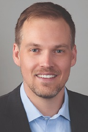
In this interview, AZoM speaks to Arno Merkle, Product Marketing Director for the TESCAN micro-CT business division.
Micro-CT is one of the most exciting areas in materials imaging. Can you explain how it works and why this is?
Micro-CT utilizes X-rays to take 2D images (radiographs) as a sample rotates 360 degrees. These projection images are then taken through a computational or ‘reconstruction’ step to form a digital 3D representation of the sample. The same principle is used for a medical CAT-Scan, but micro-CT provides much higher resolution. Part of the reason this technique has become so interesting for materials research is its non-destructive nature – enabling one to visualize and perform analysis of the interior of a sample in 3 dimensions without the need to modify or cut it. Along with unique new insights into the internal structure of samples in static states, this technique also opens doors to performing time-dependent (or ‘4D’) imaging where researchers can understand what happens inside samples as they undergo change over time.
TESCAN micro-CT specializes in dynamic or 4D micro-CT. What is dynamic-CT and what are the advantages over conventional micro-CT?
The extension of ‘static’ 3D micro-CT imaging into the time-resolved domain can be characterized by several terms, for example, time-lapse or ‘4D’ imaging. These techniques seek to image how a sample changes over a period of time, be it seconds, minutes, hours, or even days or weeks, either as an interrupted or continuous process. At TESCAN, we have developed methods that leverage our hardware and software developments to facilitate such time-dependent studies more easily, expanding the scope of the possible experiments that can be conducted in the lab.
‘Dynamic’ CT, in particular, refers to the most advanced sub-set of time-resolved X-ray imaging, where a sample is imaged continuously as it is changing. The difference between ‘dynamic’ and ‘time-lapse’ can be thought of as the difference between a smooth motion picture and a stop motion animation. The advantage of performing continuous acquisition (dynamic CT) lies in the ability to perform uninterrupted, real in situ experiments. A material undergoing change cannot always be predicted, and by utilizing dynamic CT we can now more accurately understand these processes. Up until now, the majority of 4D studies on lab-based systems were only performed using an interrupted, time-lapse methodology, while the much faster, uninterrupted, dynamic CT method had been relegated to CT beamlines at synchrotron facilities. TESCAN is focused on providing dynamic CT capability into the lab.
For those interested in a deeper dive, we recently published a paper highlighting our approach to dynamic CT. available here: DEWANCKELE, J., et al. (2020), Innovations in laboratory‐based dynamic micro‐CT to accelerate in situ research. Journal of Microscopy, 277: 197-209. doi:10.1111/jmi.12879 .
What features of your systems allow you to perform these dynamic/time-lapse studies?
A number of developments and features are required to come together in harmony to facilitate dynamic CT workflows, including hardware, software, and application workflows. On the hardware side, highly optimized components prioritize throughput, including high-power sources and efficient detectors. Other features include the ability to scan continuously without stopping every 360 degrees – this comes about by integrating dedicated rotation stage connections or, alternatively, via the unique gantry-based design of our DynaTOM where a stationary sample is imaged by a rotating source and detector. This lets researchers design in situ stages without the worry or complexity of tangling cables, fluid lines, or sensors, which are often major obstacles in a traditional geometry.
Hardware elements are truly only part of the story of what enables dynamic CT. Our developments in software and application workflows are diverse and focus on optimizing scans for continuous acquisition, reconstruction and to make visualization easier and more straightforward to the user. In all of this, we maintain a high degree of flexibility for the most complex experiments as well. Finally, the skills of our application scientists who have years of experience pioneering many dynamic imaging applications, form a world-class training and consultation team for our customers, who are never alone in their efforts.
Your systems have been optimized for dynamic performance. Does this mean you have had to sacrifice spatial resolution?
There are always trade-offs regarding speed, signal/noise, and resolution. This is true of every measuring or imaging system. We’ve developed a nice balance amongst these factors while focusing on the ability to perform dynamic CT. There are systems on the market with higher spatial resolution, but they address different needs and are incapable of performing the high speed, high fidelity, acquisitions required for dynamic CT. We view these approaches as highly complementary to one another, much like SEM, FIB-SEM, and TEM all complement one another in the field of electron microscopy.
There are many potential applications in materials science for dynamic CT, can you give us some examples?
While the list of types of in situ experiments is truly endless, we have seen intriguing uses of dynamic CT for a number of use cases in Materials Science just in the past few years. For example, mechanical testing of structural materials (typically metals, 3D printed parts or composites) is a rather common area of investigation, imaging, and analyzing how failures under load can be related to cracks, voids, and other defects. Here, we typically would image a sample as it is being compressed, bent, or placed under tension and try to correlate the deformation events with force-displacement measurements. At the point of failure or deformation, we may then perform further investigations using volume-of-interest scanning in the micro-CT or a complementary technique, such as SEM or plasma FIB-SEM to investigate finer microstructural details.
Other applications include studying the deformation of highly porous and lightweight structures, such as metal or polymer foams or even applications in food science, such as bread baking or beer foam collapse. Recently, we have been studying particle suspensions in liquid and exploring related flow behavior. Other applications include hydration, filtration, or absorption processes in various materials including consumer products. Corrosion and charge cycling of batteries represent two other very common applications, albeit on slightly longer time scales.
Most importantly, we have the ability to adapt our approach to the time-scales of interest, from a maximum tomographic resolution of <10 seconds for the fastest dynamic CT acquisitions to weeks or months if studying slow-moving processes in an interrupted time-lapse fashion, and everything in between.
Earth science is another discipline that has benefitted from the technology. Can you give us some examples of applications they are interested in?
Whether in sub-fields such as environmental geology, engineering geology, natural resources, petroleum engineering, or geodynamics, we have encountered many attractive applications that make use of dynamic CT. Examples include fracture dynamics of rock under compressive load, two- and three-phase flow in porous rock, compaction in sediments, and mineralization/dissolution processes. Most exciting is the ability to visualize and analyze the change in non-stable multi-phase flow. Without access to dynamic CT, researchers are limited to imaging liquids in an equilibrium state. In other words, they won’t actually see what is happening as different liquids initially interact with each other, only the aftermath of that interaction.
Earth Science: In situ two-phase flow in rock
Prior to being acquired by TESCAN, XRE NV had amassed many years of expertise in micro-CT imaging, building bespoke systems. This has given you an excellent insight into the architecture of these systems. Can you share with us some of the key design principles that have allowed you to build instruments suited to these types of experiments?
The most recognized unique system we have developed is the DynaTOM, with its rotating gantry-based design. This was designed from the ground up to meet the needs of in situ experiments by avoiding sample rotation, optimizing acquisition times (down to < 10 seconds), and maintaining sufficient spatial resolution. Beyond the DynaTOM’s unique design, all of our systems have been built with robustness and flexibility in mind, including stage accuracy and a high number of degrees of freedom, which enable countless acquisition geometries. We’ve also built our systems in a way to anticipate the need to communicate and adapt with third-party devices, whether in the form of an in situ device or future detectors. For power users, gaining access to control the system and its peripheral items through our acquisition software offers nearly limitless opportunities. We’ve balanced this by making our systems user friendly for occasional non-expert users.
From an operational and analytical perspective, the user interface and software play a crucial role in bringing the massive datasets together. What range of functions does your software allow and how flexible is it?
Our range of software platforms offers complete control of our micro-CT systems to conduct acquisition, reconstruction, and essential visualization for both 3D and 4D datasets. We feel strongly that both 3D and 4D workflows be served in a common environment and that links between acquisition, reconstruction, and visualization enable a better user experience. For example, we are able to conduct a fast survey acquisition, reconstruct and visualize quickly, then identify (visually) sub-volumes of interest for follow-up scans at higher resolution or detail. Once those regions are marked, the SW can send the coordinates back to our acquisition interfaces and seamlessly conduct the acquisition at the desired regions in a highly integrated manner. We call this method ‘VOIS’ for volume-of-interest scanning and believe we’ve built this in the most intuitive way, unmatched by other commercial solutions. We also have tools for easily setting up complex dynamic acquisitions, for processing dynamic CT datasets, and for visualizing them in a straightforward manner.
The only other way to carry out the types of dynamic CT experiments that your systems allow is at a synchrotron. Given access to such facilities can be difficult, do you see your systems as an effective screening tool for beamline experiments?
Synchrotron beamlines offer a fantastic breadth of capabilities and extend well beyond what laboratory systems can accomplish in many areas. However, access to these facilities is limited. Often researchers can only access this technology 1 or 2 times a year. We strongly believe that by utilizing a similar experimental approach with dynamic CT in the laboratory, researchers can optimize an experimental workflow prior to their highly valuable beamtime. We expect this symbiotic relationship between laboratory systems and synchrotrons to only grow in the future.
You have also done some more “general interest” studies that effectively showcase the applicability of dynamic CT. Can you tell us a bit about some of these?
Certain ‘general interest’ studies have gotten some attention due to the relatability of their subjects. In fact, each of these fields has strong scientific foundations and communities behind them.
For example, in Plant Science, we have studied the early germination process of cress seeds as seen in the video link below. The emergence of a radical in soil, as well as the sprouting of leaves, only scratches the surface of what we hope to explore in plant sciences:
Dynamic imaging of germinating seeds
Nearly everyone loves a tasty ice cream cone on a hot summer day. In that spirit, we imaged a melting ice cream cone in the DynaTOM, where changes can easily be witnessed. This represents only one of many examples we’ve explored within the Food Sciences, where changes in texture and structure have a direct impact on product quality and the consumer’s taste experience.
Food Science: Dynamic melting process
Lastly, the movie below highlights a vitamin pill dissolving in water, something many people can relate to. This is representative of a whole class of drug-delivery related research fields that can be conducted with dynamic CT in the pharmaceutical industry. Studying the interaction between pills and liquid environments are a key step in controlling their efficacy and optimizing their performance.
Pharmaceutical: Multi-vitamin pill dissolution
Where can people find out more information about your unique dynamic CT systems?
Come visit our website or visit our YouTube channel, where you can explore the latest in dynamic CT and learn more about our micro-CT products. You can also reach out to your local TESCAN representative to find out more information.
About Arno Merkle
 Arno Merkle directs global business development activities for TESCAN’s micro-CT product line. He joined TESCAN in early 2018, through the acquisition of Belgian-based micro-CT technology company, XRE. Prior to TESCAN, Arno spent 10+ years in the electron- and X-ray microscopy industries, serving primarily within segment and product marketing leadership roles. He received his Ph.D. from Northwestern University (Evanston, IL USA) in Materials Science and Engineering and his undergraduate B.A. in Physics at Gustavus Adolphus College (St. Peter, MN USA).
Disclaimer: The views expressed here are those of the interviewee and do not necessarily represent the views of AZoM.com Limited (T/A) AZoNetwork, the owner and operator of this website. This disclaimer forms part of the Terms and Conditions of use of this website.