This article considers the use of X-ray spectroscopy and synchrotron radiation to characterize the structure of halide perovskites.
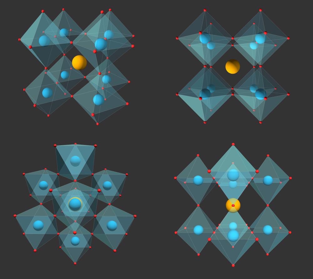
Image Credit: Niethammer Zoltan/Shutterstock.com
Perovskites have been hailed as something of a ‘wonder material’ for photovoltaic devices. This is in part due to perovskite-based devices managing to achieve efficiencies for solar energy conversion of over 20% in less than half a decade of research – a rate of improvement that no other material has seen.
The term perovskite refers to the particular crystal structure of this family of materials. Their atomic composition can vary, with both organic and inorganic variants of the material in existence.
Tuning the optoelectronic properties of perovskite can be done through doping and changing the morphology of the structure.
The sensitivity of the performance of perovskite materials to both chemical composition and morphology means that any characterization of the materials requires a technique with high chemical sensitivity and spatial resolution.
X-Ray Spectroscopies
X-ray spectroscopies are usually the tool of choice for materials characterization. This includes techniques such as X-ray powder diffraction, where a crystalline sample is irradiated with X-rays and a moveable detector records the intensity of the diffracted radiation at different positions.
The intensity patterns can then be used to reconstruct information about the atomic arrangement of the material.
However, recording X-ray spectra of halide perovskites is challenging, as the beam intensities can result in sample damage. Achieving high spatial resolution in techniques such as X-ray microscopy requires the use of tightly focused beams which in turn puts a high thermal load on the sample.
The soft nature of halide perovskite films also makes the issue of other electron microscopy techniques, such as transmission electron microscopy (TEM) for characterization of their structure and composition, problematic as the focused ion beams tend to induce changes in the sample before measurements can be performed.
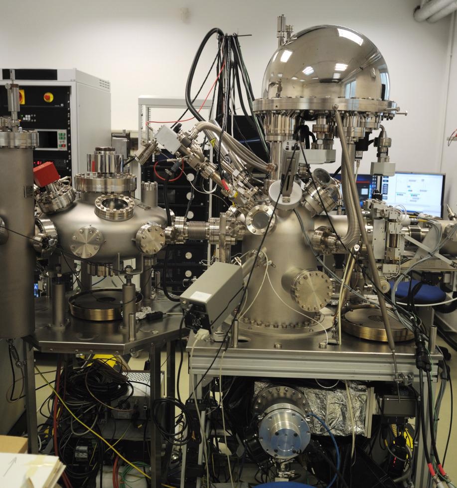
X-ray photoelectron spectrometer. Image Credit: Tomas Salkus/Shutterstock.com
With X-ray spectroscopies, however, particularly with highly sensitive detectors, it is possible to work under sufficiently low irradiation conditions so that the perovskites can be measured in the same state that they would be present in in an operating device.
While care needs to be taken to avoid damage conditions and ensure the measurement process does not influence the outcome, there are a number of methods compatible with the measurement of halide perovskite thin films.
Synchrotron Radiation
Synchrotrons are advanced light sources capable of generating X-ray radiation with high spatial coherence, excellent brightness and with wide energy tunability. This makes them an ideal photon source for performing a variety of measurements for the characterization of halide perovskites.
The high brightness means that even weak diffraction signals can be detected and better quality scattering patterns can be obtained.
Depending on the capabilities of the synchrotron facility, a single light source can be used to generate photons of energies spanning from the ultraviolet range through to the hard X-ray.
One approach to elemental characterization is to use high energy radiation to ionize the core electrons in a sample.
These are the most tightly bound electrons, closest to the nucleus, and even in complex molecular species the core electrons have energies that are characteristic of the element they are removed from and can be used for elemental analysis of the material.
The core electrons of light elements are less tightly bound than those of the heavier elements. What this means is most organic elements, like carbon, nitrogen and oxygen, can be core ionized with energies less than 1 keV – or soft X-rays.
Elements such as lead and metals require significantly higher energy electrons. Few other light sources can produce photons over such a great energy range, and this is particularly useful for halide perovskite analysis as they can contain a mixture of organic and inorganic elements.
Structural Sight
For halide perovskites, synchrotron radiation sources have been used to reveal a wealth of information about the structure and function of such materials. This includes investigating how the degree of order in the crystalline structures affects the device performance and the still poorly understood role that grain boundaries play.
Doped structures can also have some degree of ion mobility within the crystalline lattice, and so X-ray experiments performed with synchrotron radiation can be used to identify the degree of doping, the site in the structure the dopant is present, and how this may migrate through the structure.
X-ray measurements of halide perovskites have been moving from isolated thin films to true in operando measurements on full devices. This has included looking at changes in the structure while devices are under thermal, electrical or pressure loads, and capturing what degradation processes occur after long operating times.
One of the biggest current challenges in making full use of the suite of multimodal X-ray techniques available for halide perovskite characterization is dealing with the challenge of increasingly large datasets and finding automated ways to perform this analysis online during experiments.
Given the issues with the degradation of such material, moving towards shorter measurement times under less intense radiation conditions will also be highly beneficial for future measurements.
References and Further Reading
Ansari, M. I. H., Qurashi, A., & Nazeeruddin, M. K. (2018). Frontiers, opportunities, and challenges in perovskite solar cells: A critical review. Journal of Photochemistry and Photobiology C: Photochemistry Reviews, 35, 1–24. https://doi.org/10.1016/j.jphotochemrev.2017.11.002
Qiu, T., Hu, Y., Xu, F., Yan, Z., Bai, F., Jia, G., & Zhang, S. (2018). Recent advances in one-dimensional halide perovskites for optoelectronic applications. Nanoscale, 10(45), 20963–20989. https://doi.org/10.1039/c8nr05862h
Stuckelberger, M. E., Nietzold, T., West, B. M., Luo, Y., Li, X., Werner, J., Niesen, B., Ballif, C., Rose, V., Fenning, D. P., & Bertoni, M. I. (2020). Effects of X-rays on Perovskite Solar Cells. Journal of Physical Chemistry C, 124(33), 17949–17956. https://doi.org/10.1021/acs.jpcc.0c04645
Zhou, Y., Zhou, H., Deng, J., Cha, W., & Cai, Z. (2020). Decisive Structural and Functional Characterization of Halide Perovskites with Synchrotron. Matter, 2(2), 360–377. https://doi.org/10.1016/j.matt.2019.12.027
Disclaimer: The views expressed here are those of the author expressed in their private capacity and do not necessarily represent the views of AZoM.com Limited T/A AZoNetwork the owner and operator of this website. This disclaimer forms part of the Terms and conditions of use of this website.