In this interview, AZoM talks to Ron Rubinovitz from Thermo Fisher Scientific about the utilization of FTIR microscopy for automated microparticle analysis.
Please can you introduce yourself and automated microparticle analysis by FTIR microscopy?
Hello, my name is Ron Rubinovitz, and I will discuss automated microparticle analysis by Fourier transform infrared (FTIR) microscopy. My goal today is to show that analyzing hundreds of particles on a surface can be straightforward and routine with the right tools.
What are the benefits of FTIR spectroscopy and IR microscopy?
Infrared (IR) microscopy, like typical FTIR, gives the user a rapid, non-destructive characterization technique. Although the infrared spectrum gives important insights into the molecular structure of the sample through the location and shapes of infrared absorption peaks, more can identify samples through the use of available extensive IR spectral libraries.
Just like macro FTIR measurements, FTIR microscopy can use different collection techniques depending on what is optimal for the sample. These collection modes are transmission, reflection, or attenuated total reflections (ATR).
The advantage of IR microscopy is that samples that would be too small or difficult to handle can be analyzed relatively straightforwardly. Similarly, larger samples of interest in how the composition changes across the sample are often also analyzed by FTIR microscopy.
Please can you tell us about the Nicolet RaptIR FTIR Microscope and its features?
First, I want to point out a massive development incorporated into the new microscope, Thermo Scientific™ OMNIC™ Paradigm Software. This is a true 64-bit software that allows many advanced features that were never part of our previous Thermo Scientific™ OMNIC™ Software. At the same time, Paradigm can read legacy OMNIC spectra libraries and maps.
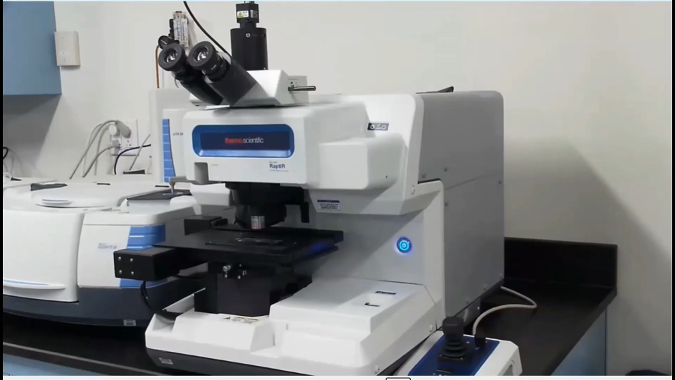
Image credit: Thermo Scientific - Chemical Analysis
The OMNIC Paradigm Software increases the capabilities of the Thermo Scientific™ Nicolet™ RaptIR™ FTIR Microscope. Multiple collections can be set up and run automatically without interruption. We can combine various sample points, line maps, or area map collections.
Another innovation of the Nicolet RaptIR is integrating a 4x glass lower magnification objective with a 15x IR objective. Mosaic images from each objective are automatically collected and superimposed onto each other. This makes it much easier to find specific regions of interest in larger samples and start collecting spectra faster.
We have also improved the stage so it has a 40-millimeter working distance. This provides much more flexibility in working with larger samples, such as environmental cells or large geologic samples.
Novel mapping features of the Nicolet RaptIR microscope make collecting maps quicker and more efficient. The high-speed collection mode allows the collection of 10 scans per second, and the OMNIC Paradigm Software allows you to check the values for aperture, step size, number of scans, etc, before or after the collection of one measurement or even multiple maps.
Many automated features have been added to increase sample collection's ease, efficiency, and reproducibility. First, the user can dial in the specific pressure between the ATR crystal and the sample when doing ATR measurements. We also have automated switching between the 4x glass and 15x IR objectives. This is particularly useful when trying to find a small section of a sample you are interested in.
Software-controlled visible and IR polarizers are available to provide insight into the molecular structure of samples. Users will also quickly notice that the stage movements are faster, making adding or removing samples easier, for example.
Why is the RaptIR applicable to use in the pharmaceutical industry?
Instrument performance verification is important to everyone and critical in the pharmaceutical world. We have software methods for automated performance verification and specific pharmacopeia tests using a performance checkpoint. Pharmaceutical customers will want to use the traceable certified check plate. The Nicolet RaptIR FTIR Microscope comes with a performance check plate.
What is automated particle analysis and where is it used?
The identification of unexpected particles is an essential task in many laboratories. For example, in the world of pharmaceuticals, the presence of particles in injectables is an area of extreme concern. For scientists studying the environment, identifying microplastics in otherwise natural settings is also an area of intense interest.
Similarly, the routine and efficient identification of important particles is vital in manufacturing processes. Particle ID provides the key to the origin of particles. For example, in the manufacturing world, the composition of unexpected particles can provide clues as to whether they are from an unanticipated pathway in the process or small external particulates from a degrading gasket.
For environmental scientists, the source of microplastics is often determined by the type of polymer that has been found. In addition to the chemical composition, the shape of the particle can provide information about its origin.
The challenge in these measurements has typically been that the number of particles involved can be high enough to make manual targeting and analysis of particles difficult and time-consuming. This step can be done remarkably well in an automated fashion by the OMNIC Paradigm Software.
The focus of this interview is the actual collection and identification of particles, but I also want to say a quick word about sample presentation, mainly when particles are found in a liquid solution. For example, this can be the case in pharmaceutical manufacturing or waterworks facilities.
Most often, these measurements are done most effectively in reflectance mode.
Why are ATRs or transmission modes not typically our first choice when measuring many particles simultaneously?
ATR of multiple particles can result in the particles sticking to the crystal and being accidentally measured repeatedly. The transmission mode is somewhat limiting since the particles must be transferred to an IR window.
However, using a filter that is a reasonably good reflector, particles from a liquid can be isolated and brought directly to the microscope. Different methods and materials can be used to isolate the particles for IR analysis. For example, a solution could be filtered through a silicon filter. The filter can then be mounted in a holder for reflectance measurement on the Nicolet RaptIR FTIR Microscope.
Can you give us an overview of the Nicolet RaptIR FTIR Microscope workflow applied to automated particle analysis?
In this example workflow for automated particle analysis, you would have the Nicolet RaptIR microscope, including both eyepiece, video camera and a computer-controlled stage, connected to the research grade Thermo Scientific™ Nicolet™ iS50 FTIR Spectrometer.
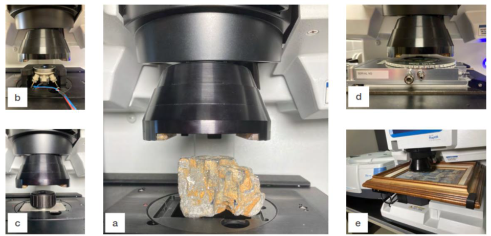
Image credit: Thermo Scientific - Chemical Analysis
Imagine you have a silicon filter mounted onto a glass side. The sample is mounted on the stage with particles below the 4x glass objective. Then, you switch to the OMNIC Paradigm Software to start configuring the measurement.
Various parameters are visible on the system. You can autofocus the sample before capturing any video if wanted. You can put the session name in. You also have other parameters that can be associated with your collection, so these are absorbents, percent transmittance, peaks up, peaks down, the number of scans, whether the mosaic is going to be collected at the current location on the stage, do we want it to run over the entire slide or just the center of the glass slide, etc.
It can also allow for the auto illumination check in automatic mode, or we can guide it on our samples' nature. On the other side, I could choose the resolution. The collection mode can be set at transmission, reflection, or ATR.
We also have step size, aperture height and aperture width. In the example of particle analysis, the system will figure out the step size, the aperture height, and the aperture width appropriate for every particle it finds.
After naming the session and giving it a tag, the important features are now set and, upon clicking “start session”, you can move forward.
The Nicolet RaptIR will collect an image of the sample. You can then select the region within that video capture you want to analyze and guide the software in defining the particles on the filter. Once clicking start, the software quickly changes to a microscope setup tab, performing an auto illumination.
Then it progresses to using the glass objective to collect a mosaic. The silicon filter is mounted in its slide mount, and many filter particles sit on top. At this point, the system automatically switches between the glass and IR objectives. At that point, auto illumination and autofocus will occur again for this objective.
We do not have to live and die by the Paradigm choice of best focus, best illumination. We can always override that if we would like, but I find it almost always gets you in a good position, and you can always adjust it as needed. Once this is done, you can go to the camera view and a red square will show your location.
The joystick can be used to move around the surface. Within this camera view, there are a lot of other controls. For example, if I want to change the illumination, there is a slider bar or the opportunity to type numbers in for focus and illumination. If you find yourself running the same type of sample day in and day out, you can take advantage of that.
Then, you can select ‘particle analysis’ and draw a square or rectangle over the sample. The software will then switch over and analyze how many particles are within that area. The area can be adjusted easily.
After accepting its particle calculation, it returns to the microscope setup cam to start the actual collection of spectra. Using the information from the captured image of the particles on the filter, the OMNIC Paradigm Software will collect the necessary background scans of the required aperture settings in your selected location. The OMNIC Paradigm Software will begin the collection of individual particles using the locations determined in the previous step.
After this, you can choose the location for the background. You could measure the background, but in particle analysis mode, it is quicker to click on the sample.
The system then runs each of those apertures and decides for whatever number of particles that I told it to do that it is going to run, for example, eight apertures. It has eight unique aperture settings that it needs to collect in that mode. Once it is done with that, you will see that the stage starts moving after collecting each particle. After a quick collection, the spectra show below and the system moves on to the next particle.
As it changes to each particle position, the spectrum below will update and you can leave the camera mode on if you want.
If you want to verify what has happened, you can zoom in and check to see what each spectrum looks like. One of the options is sending this spectrum to the database, so you could take each spectrum, store it and analyze it, but this would be pretty tedious. It is more efficient to take advantage of the automation.
The analysis of the particles and the OMNIC Paradigm Software will produce tables showing the identification of different materials and calculating what percent of the particles are represented. You will have a row associated with every particle you have measured with information, such as its identification, its identification match value, and dimensional information.
Within the system, there is a listing of all the measured particles. You can drive the aperture information and the collection time for each one of those particles. All that information is stored in the file in our database.
The next thing you could do is identify these particles automatically. You can go to the search setup, verify that you have the right threshold and library, and analyze these over the full spectral range.
The system considers all these particles and identifies them. It gives a table of component names and, for example, how many particles of each type and what color they are colored in. This table shows lots of information on each particle, such as its area and circularity.
The system can also show us plots like particle distribution for an identified substance by area. It gives options for distribution, perimeter, circularity, and maximum and minimum projection. This can be done for each particle.
The system can also create a report. This makes it easy to share the information with others. This can be printed, saved to the database or even saved in Excel format.
Please can you summarize the benefits of the Nicolet RaptIR FTIR Microscope for automated particle analysis?
The Nicolet RaptIR has the highest quality infrared and visible microscope resolution. It accommodates large and heavy samples with ease. Image-centric software and hardware make finding your sample and collecting maps easier, stringing together a series of maps without stopping between collections.
There is improved automation such as ATR control - the ATR contact between the crystal and the sample can be controlled precisely and reproducibly. We have got control over the infrared and visible polarizers and, of course, we have automation that helps in our collection of microparticles.
Using the Nicolet RaptIR particle analysis function, you can take advantage of each particle's automatic location and measurement. Each particle is automatically assigned its own optimized aperture settings. The collection is fast as no time is wasted collecting areas of filter that do not have particles. ID and dimensional information for each particle as well as the sample as a whole can then be compiled into a report.
About Ron Rubinovitz
Ron earned his B.S. in Chemistry at Brandeis University and his Ph.D. in Physical Chemistry at the University of Pennsylvania where he focused on optical spectroscopic techniques. Ron then went onto a postdoctoral position at the Naval Research Laboratory utilizing infrared spectroscopic methods to analyze thin films.
Ron is currently a Senior Application Scientist for the Nicolet FTIR spectroscopy product line at Thermo Fisher Scientific and operates an applications lab in Wilmington, DE. He works with FTIR, FTIR-microscopy, chemometrics and FT-Raman spectroscopy.
This information has been sourced, reviewed and adapted from materials provided by Thermo Fisher Scientific – Chemical Analysis.
For more information on this source, please visit Thermo Fisher Scientific – Chemical Analysis.
Disclaimer: The views expressed here are those of the interviewee and do not necessarily represent the views of AZoM.com Limited (T/A) AZoNetwork, the owner and operator of this website. This disclaimer forms part of the Terms and Conditions of use of this website.