The UVM-1TM is an ultraviolet microscope with visible and near-infrared imaging capabilities. This UV-visible-NIR microscope combines state-of-the-art UV, color, and NIR imaging and viewing technologies with sophisticated optics and Scorpii Advanced Lighting Systems. The microscope has an adaptable architecture that is incredibly user-friendly and robust. Modern CRAIC optics are used in its design to provide the best image quality and many years of use.
With only one microscope and no need to switch out parts, the UVM-1TM UV microscope can image transmission, reflectance, polarization, and even fluorescence from the UV, visible, and NIR regions with great spatial resolution.
Below are microscopic samples are imaged using UV, optical, and near-infrared on the UVM-1™ microscope.
NIR Transmission Image of an integrated circuit using the UVM-1™ Microscope
Image Credit: CRAIC Technologies
Mouse tongue at 280 nm following radiation exposure. This transmission image shows the damage to the cellular structure.
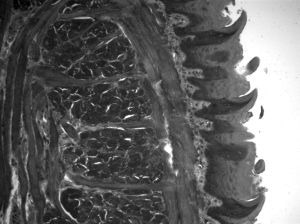
Image Credit: CRAIC Technologies
UVM-1 UV-visible-NIR Microscope
Video Credit: CRAIC Technologies
Key Features
- In-depth ultraviolet microscopy
- Vibrant microscopy
- NIR microscopy—all without switching lenses and using the same instrument
- Optional Raman analysis of small-scale specimens.
- Imaging of microscopic samples using transmission, reflectance, fluorescence, and polarization can all be done with the same tool.
- Direct vision with eyepieces as well as true high-quality digital imaging
- Simple to operate and keep up
More Capabilities
- Raman analysis of small-scale specimens
- UV, visible, and infrared spectroscopy of small samples
- Hardware, such as automation and specialist software for analysis
- Software for imaging, databasing, and specialist data analysis is included
Applications
- Analysis of cellular structure
- Crystals of proteins
- Microspectroscopy of plankton
- Tissue examination
- Microspectroscopy in vivo
- Microspectroscopy in vitro
- spectroscopy of kinetics
- Chemistry in combinatorial form
- Medication development