Nanoporous gold research has gathered momentum in the last two decades, with the material demonstrating promise in numerous bioscience, sensing, and catalysis applications, as well as for energy storage applications.1,2
Precise nanoporous gold characterization is critical for the continued development of these technologies.
Research conducted by Fraunhofer IFAM demonstrates how dual-beam Focused Ion Beam, and Scanning Electron Microscopy (FIB-SEM) imaging techniques can be used with physical modeling, advanced image-processing, and Pore Network Modeling (PNM) to supply thorough quantitative analyses of nanoporous gold samples.
Nanoporous Gold contains pores with diameters of the order of tens of nanometers, the precise size of which can effortlessly be tuned during fabrication.
The subsequent biocompatible, physically stable, and nanotextured surface have made it particularly appealing for drug release, biosensing, and selective catalysis applications.3–5 Other notable properties include high electrical conductivity and plasmonic properties. 6,7
It is easy to modify the surface of nanoporous gold by utilizing self-assembled monolayer (SAM) technology, or it can simply adsorb certain molecules directly. Tuning the pores to the same order of size as antibodies, proteins, enzymes, and oligonucleotides can enable the incorporation of such biomolecules within the structure to create biosensors.
Likewise, tuning the pore size to the order of specific chemical reactants has been observed to produce reusable and very specific catalysts. The same porous structure and high surface area have also led to an investigation into nanoporous gold’s potential applications in fuel cells and supercapacitors.8,9
Imaging and Analyzing Nanoporous Gold
The morphology of the porous structure of prepared nanoporous gold samples is very changeable, and the samples’ performance and behavior in the applications mentioned above are predominantly decided by their pore formation.
Their characterization is dependent upon the size, shape, and distribution of pores and potentially the channels which connect them. The advancement of imaging-based techniques that are able to quantify porosity and its flow properties, such as permeability and tortuosity, is critical in order to develop nanoporous gold-based technology.
Dr. K Thiel of Franhofer IFAM has demonstrated that a complete quantitative analysis of nanoporous gold samples can be accomplished through utilizing FIB-SEM imaging and subsequent image analysis.
Employing a Thermo Scientific™ Helios DualBeam system for FIB-SEM imaging ensures it is possible to gather high-resolution images and, at the same time, ablate or “mill” the surface to uncover internal pore structures.10,11
This technique can be used to help optimize nanoporous fabrication for various applications. The nanoporous gold sample was imaged using a Helios DualBeam. The voxel size of the last image is about 10x13x10 nm, and the overall imaged volume is about 10x11x10.41 µm.
The obtained images were processed using Thermo Scientific™ Avizo™ Software (Figure 1). In the first instance, Avizo Software enabled the removal of jitter and misalignment artifacts, which frequently occur in FIB-SEM imaging techniques.
The alignment algorithm can automatically adjust for translational and rotational misalignments based on the similarity of sum-of-squared-differences images.
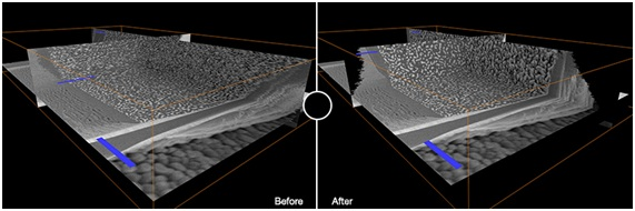
Figure 1. FIBSEM images of nanoporous gold obtained by Thermo Scientific Helios DualBeam before and after automatic alignment done with Avizo Software. Sample courtesy of K.R. Mangipudi, Institute for Materials Physics, University of Goettingen.
Helios DualBeam has offered high-quality images with limited noise and no pore-back effects from open pores, removing the requirement for additional processing.
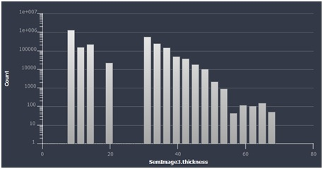
But beyond simply aligning the 2D stack of FIB-SEM images, Avizo Software computes a number of the key statistics from a 3D dataset that defines the porosity of the sample.12
These include the pore morphology and connectivity, together with fragmentation index, volume fraction, average thickness, and average spacing of pores.
It would also be possible to use the resultant 3D model of the sample for physical modeling, including calculating the permeability tensor and the sample’s absolute permeability.
Pore Network Modeling
Most crucially, Avizo Software also allows Pore Network Modeling (PNM), in which complex porosity networks can be estimated to precisely predict the flow properties inside the porous structure.
The analysis is conducted on a 3D SEM data set and allows the quantities such as the total number of pores and throats in the sample to be calculated, as well as their volume, equivalent radius, and coordination number.
This data was then utilized to model the sample’s flow characteristics, calculating precise values for flow rate, permeability, and tortuosity.
Bearing in mind the significance of the porous structure in catalysis and sensing applications, these metrics can be employed to evaluate material performance and thus inform the manufacturing process of nanoporous.
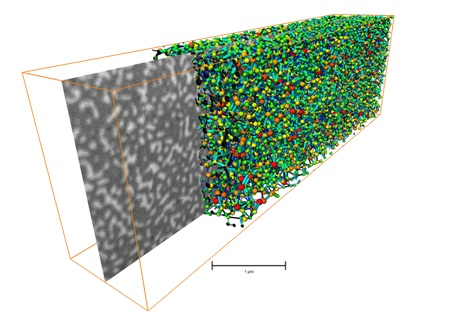
Figure 2. Representation of the pore space using a spheres and tubes model.
Avizo Software for Porosity Analysis
Avizo Software is a self-contained analysis platform that includes quite a few specialized features for the analysis of porous materials.
Avizo Software is compatible with a variety of imaging methods including microCT, SEM, FIB-SEM, and TEM, which facilitates the quantification of micropores, large macro-voids, sponge-type voids, and inclusions.
The convenience of procuring visually intuitive 3D representations of a sample with detailed statistical analysis and state-of-the-art physical modeling provides researchers with a simple method of acquiring comprehensive and thorough characterizations of porous materials such as nanoporous gold.
Pore Network Modeling provides a practical and efficient way of ascertaining the performance of nanoporous gold and other porous materials for biosensing, catalysis, and additional applications.
Avizo Software is also specialized for multiple other applications in bioscience, nanotechnology, and materials research. Such applications include the characterization of fibers in composite materials and internal strain mapping.
Wherever quantitative analysis of micro- and macrostructures of materials is necessary, Avizo Software provides an all-in-one solution.
References and Further Reading
- Stine, K. J. Nanoporous gold and other related materials. Nanomaterials 9, (2019).
- Detsi, E., Chen, Z. G., Vellinga, W. P., Onck, P. R. & De Hosson, J. T. M. Actuating and sensing properties of nanoporous gold. J. Nanosci. Nanotechnol. 12, 4951–4955 (2012).
- Bhattarai, J. K. et al. Preparation, modification, characterization, and biosensing application of nanoporous gold using electrochemical techniques. Nanomaterials 8, (2018).
- Seker, E., Berdichevsky, Y., Staley, K. J. & Yarmush, M. L. Microfabrication-compatible nanoporous gold foams as biomaterials for drug delivery. Adv. Healthc. Mater. 1, 172–176 (2012).
- Yan, M. et al. Nanoporous Gold Catalyst for Highly Selective Semihydrogenation of Alkynes: Remarkable Effect of Amine Additives. J. Am. Chem. Soc. 134, 17536–17542 (2012).
- Chen, L. Y. et al. Toward the Theoretical Capacitance of RuO 2 Reinforced by Highly Conductive Nanoporous Gold. Adv. Energy Mater. 3, 851–856 (2013).
- Maaroof, A. I., Gentle, A., Smith, G. B. & Cortie, M. B. Bulk and surface plasmons in highly nanoporous gold films-a.
- Nagle, L. C. & Rohan, J. F. Nanoporous gold anode catalyst for direct borohydride fuel cell. in International Journal of Hydrogen Energy 36, 10319–10326 (Pergamon, 2011).
- Lee, K. U., Byun, J. Y., Shin, H. J. & Kim, S. H. A high-performance supercapacitor based on polyaniline-nanoporous gold. J. Alloys Compd. 779, 74–80 (2019).
- An Introduction to Electron Microscopy - FIB - A focused ion beam (FIB) instrument is almost identical to a SEM, but uses a beam of ions rather than electrons | Thermo Fisher Scientific. Available at: https://www.fei.com/introduction-to-electron-microscopy/fib/. (Accessed: 18th February 2020)
- Dual Beam | Dual Beam FIB | Thermo Fisher Scientific - UK. Available at: https://www.thermofisher.com/uk/en/home/industrial/electron-microscopy/electron-microscopy-instruments-workflow-solutions/dualbeam-microscopes.html. (Accessed: 18th February 2020)
- Avizo for Porosity Analysis | Thermo Fisher Scientific - UK. Available at: https://www.thermofisher.com/uk/en/home/industrial/electron-microscopy/electron-microscopy-instruments-workflow-solutions/3d-visualization-analysis-software/avizo-porosity-analysis.html. (Accessed: 18th February 2020)

This information has been sourced, reviewed and adapted from materials provided by Thermo Fisher Scientific.
For more information on this source, please visit Thermo Fisher Scientific – Electron Microscopy Solutions.