In this interview, industry experts Matt Joens and Jaspreet Singh explain how FIB-SEM and FTIR can be combined to isolate, extract, and identify small organic contaminants in complex samples.
To get started, can you explain why a combined FIB-SEM and FTIR workflow is useful for analyzing organic contaminants?
Matt Joens: A combined FIB-SEM and FTIR workflow is useful because many contamination challenges are becoming smaller, more complex, and harder to access using traditional preparation methods.
In many cases, contaminants are embedded within a matrix or located in areas that are difficult to reach manually. This is especially relevant for microplastics, manufacturing residues, medical devices, semiconductor devices, and pharmaceutical samples.
The FIB-SEM workflow allows us to locate, image, mill, and isolate a specific region of interest with high precision. Once the contaminant has been extracted, FTIR can provide the spectral information needed to identify the material. The SEM provides information on size, texture, morphology, and surface details, while FTIR confirms the chemical identity of the organic contaminant.
Together, the two techniques provide a more complete analytical workflow than either method could offer alone.

Image Credit: BG-Studio/Shutterstock.com
What types of applications can benefit from this workflow?
Matt Joens: There are many applications where this type of workflow can be valuable. One major area is microplastics analysis, particularly as microplastics and nanoplastics are becoming a growing concern in water, crops, food sources, and human health. These particles are increasingly found throughout the environment, so being able to track and identify them is becoming more important.
The workflow is also useful in manufacturing failure analysis. For example, if a black powder, residue, or unexpected contaminant appears during production, a sample can be collected and analyzed to determine what it is and where it may have come from.
This is important in QA and QC because fast contaminant identification can help manufacturers respond quickly, reduce disruption, and prevent wider production issues.
It can also support pollution monitoring, pharmaceutical analysis, medical device investigations, semiconductor failure analysis, and polymer-related research, especially where small organic contaminants need to be isolated and identified.
What does SEM contribute to the analysis?
Matt Joens: SEM, or scanning electron microscopy, provides high-resolution surface information that goes beyond what a light microscope can achieve. It is not a diffraction-limited tool, which means it can reveal very small surface details, including morphology, texture, and particle size.
SEM is used across many industries, including life sciences, pharmaceutical sciences, materials science, and manufacturing. Depending on the detector being used, SEM can provide different types of information.
Some detectors give strong Z contrast, which is useful for identifying metallic contaminants, while others provide more surface-sensitive information and highlight fine textural details.
SEM can also be paired with EDS or EDX to provide elemental information. This allows users to perform point, area, and line scans, as well as phase mapping.
However, when analyzing polymers or other organic materials, EDS and EDX are limited. They may show that a sample contains carbon and oxygen, but they will not identify the specific polymer or side chains. That is why FTIR is needed as a complementary technique.
What is FIB-SEM, and how does it help isolate contaminants?
Matt Joens: FIB-SEM, also known as a dual beam system, combines a scanning electron microscope with a focused ion beam. The SEM is used to observe the sample, while the focused ion beam can cut, mill, or ablate material from a very specific area. This makes it possible to isolate a contaminant from the surrounding matrix.
The FIB can also deposit material. By introducing an organometallic gas into the chamber, the beam can cleave off the organic component and leave behind a mostly metallic residue. This can be used to attach a small isolated sample to a Tungsten lift-out needle.
By combining ion milling, metal deposition, and lift-out, FIB-SEM allows very specific regions of interest to be extracted from complex samples. This is especially useful when the contaminant is too small to manipulate by hand or is embedded inside another material.
What does FTIR add to the workflow?
Jaspreet Singh: FTIR stands for Fourier transform infrared spectroscopy. It is a type of infrared spectroscopy that uses an infrared source, an interferometer, and a Fourier transform to perform rapid, non-destructive analysis.
This technique provides information on molecular structure and functional groups, enabling materials to be identified through library searching.
FTIR microscopy takes the capabilities of FTIR spectroscopy and incorporates them into a microscope. This allows smaller samples to be analyzed, while also enabling additional capabilities such as mapping and automated analyses.
Like regular FTIR spectroscopy, FTIR microscopy can be performed in different modes, including transmission, reflection, and attenuated total reflectance. In such a workflow, FTIR provides the molecular identification that SEM and FIB-SEM cannot provide alone. SEM and FIB-SEM can locate and extract the contaminant, but FTIR confirms what the material actually is.
How is FTIR microscopy used in small-scale material analysis?
Jaspreet Singh: FTIR microscopy is useful for analyzing or mapping small samples. In pharmaceutical sciences, for example, it can be used to map the surface of a tablet and show the distribution of different chemical features. These maps can be generated by correlating a spectrum with the rest of the data set, using peak area, peak height, ratios, or other spectral features.
FTIR microscopy is also widely used in microplastics analysis. Automated particle analysis can recognize particles on a filter and then analyze them individually. This makes it possible to identify different polymer particles in a sample.
Other applications include forensic analysis, such as ink analysis and automotive paint chip analysis. Across these areas, FTIR microscopy provides both visual and chemical information, helping researchers understand where a material is located and what it is.
How does the FIB-SEM to FTIR workflow work step by step?
Jaspreet Singh: The workflow begins with the identification of a contaminant in the SEM. Once the region of interest has been located, the FIB is used to isolate the material by milling away the surrounding matrix. The sample can then be lifted out using a probe and deposited onto an IR-reflective substrate for FTIR analysis.
This combination is valuable because each technique addresses a limitation of the other. FTIR may struggle to directly analyze embedded materials because the matrix can obscure the sample or because the particle may be too small to manipulate.
SEM and FIB-SEM can locate and isolate the material, but they are limited when it comes to identifying the exact organic composition. By using the two techniques together, it becomes possible to extract a small contaminant and then identify it spectroscopically.
How was the workflow tested using microplastic contamination?
Matt Joens: To test the workflow, microplastics were pressed into a microcrystalline cellulose matrix. A light microscope image was first used as a map to help navigate the sample in the SEM. Once in the SEM, the different detectors showed contrast between the cellulose and the plastic regions. The lighter material corresponded to the cellulose, while the darker regions corresponded to the plastic.
A focused ion beam was then used to mill away the front, back, and sides of the selected polymer contaminant. This removed the surrounding matrix and left behind a cleaner, more isolated plastic sample.
A tungsten lift-out needle was brought to the corner of the isolated region and attached using a platinum weld. The particle was then cut free, lifted out of the sample, and transferred onto a gold slide.
The gold slide acted as an IR-reflective substrate for FTIR analysis. This approach also meant the extracted sample could be transported if the FTIR instrument was not located in the same room, building, or site as the FIB-SEM.
What did the FTIR results show after the contaminant was extracted?
Jaspreet Singh: Before analyzing the extracted contaminant, ATR FTIR spectroscopy was used to confirm that the matrix and the contaminant had distinct spectral features. The microcrystalline cellulose matrix and the polymer microspheres produced different spectra, so the extracted material was expected to resemble the polymer microspheres spectrum.
The first analysis of the extracted sample was performed in reflection mode. The spectrum showed some noise and fairly broad peaks, and the library matches were in the high 70s for a generic plastic or polyethylene. This suggested that the sample thickness, SEM coating, or weld mark may have affected the spectral quality.
To improve the analysis, the sample was compressed. This removed the top layer and thinned the sample, which significantly improved the spectrum. After compression, the reflection mode analysis produced library matches higher than 96 to polyethylene.
A second extracted sample was also transferred onto a diamond compression cell and analyzed in transmission mode, producing high-quality spectra with matches greater than 97 to polyethylene. This confirmed that the extracted organic contaminant was polyethylene.
Where could this workflow be applied in the future?
Matt Joens: One important future application is microplastics research in human health. Microplastics have been reported in different tissues throughout the human body, and researchers are increasingly trying to understand where these particles accumulate and whether different polymers are associated with different biological outcomes.
SEM and polarized light microscopy can help locate suspected plastic particles in tissue, but FTIR can help confirm whether they are polymers and identify which polymers are present.
Jaspreet Singh: Beyond biological and environmental research, this workflow could be valuable in many industrial and academic laboratories. Failure analysis labs often need to isolate a contaminant and identify the root cause of a failure.
The medical device, semiconductor, polymer, and pharmaceutical industries frequently deal with small contaminants that are difficult to handle and require both imaging and chemical identification.
Overall, the workflow provides a more holistic answer. SEM locates and images the material, FIB isolates and extracts it, and FTIR identifies the unknown. This makes the combined workflow valuable for laboratories that need to better understand small organic contaminants in complex samples.
Watch the Full Webinar Here
About Matthew Jones

Matthew Jones is a Senior Sales Development Representative at Thermo Fisher Scientific focused on SEM and FIB-SEM technologies. He specializes in advanced imaging, contamination analysis, and complex sample preparation workflows, with experience spanning biological and materials science applications.
About Jaspreet Singh
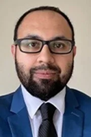
Jaspreet Singh is a Senior Application Scientist for the Nicolet FTIR spectroscopy product line at Thermo Fisher Scientific. He earned his PhD from the University of Oklahoma, where his research focused on infrared spectroscopy and advanced FTIR sampling methods.

This information has been sourced, reviewed, and adapted from materials provided by Thermo Fisher Scientific - Vibrational Spectroscopy.
For more information on this source, please visit Thermo Fisher Scientific - Vibrational Spectroscopy.
Disclaimer: The views expressed here are those of the interviewee and do not necessarily represent the views of AZoM.com Limited (T/A) AZoNetwork, the owner and operator of this website. This disclaimer forms part of the Terms and Conditions of use of this website.