In this interview, AZoMaterials speaks with Eddy Hill, Sector Manager at ZEISS Microscopy, about how advances in automated multimodal microscopy are transforming mineralogy. Eddy discusses why accurate mineralogical data is fundamental to efficient mining, explains the value of integrating light, X-ray, and electron microscopy into connected workflows, and explores how artificial intelligence is making automated mineralogical analysis more accessible, scalable, and actionable.
Can you please introduce yourself and your role at ZEISS Microscopy?
My work focuses on developing advanced microscopy solutions for geoscience and mining applications, particularly automated mineralogy and process mineralogy. I work closely with researchers and industry partners to develop workflows that combine different microscopy techniques, helping users obtain more meaningful mineralogical information and ultimately make better operational and research decisions.
Watch the full webinar here
Why is accurate mineralogy so important throughout the mining process?
Although mining operations extract rock, the objective is ultimately to recover valuable minerals and metals. The minerals themselves determine how effectively the material responds during processing, making them the reactive component throughout the recovery circuit.
Feed variability, mineral liberation, mineral associations, and the presence of deleterious phases all influence plant performance. By understanding the mineralogical characteristics of an ore body, operators can optimize grinding, improve recovery efficiency, reduce processing costs, and make better decisions throughout the operation. Process mineralogy provides the crucial link between the geology of a deposit and its metallurgical performance.
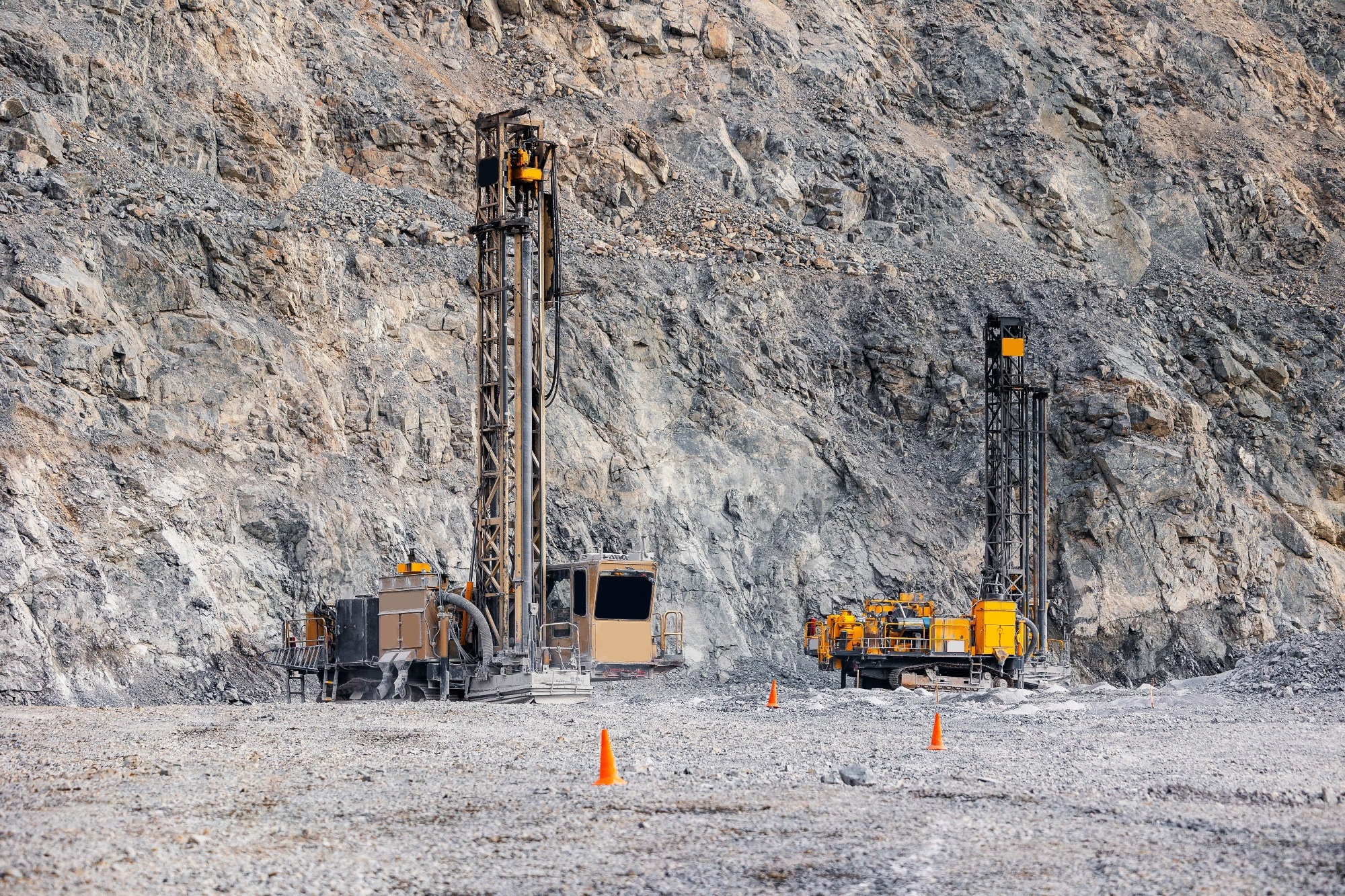
Image Credit: Parilov/Shutterstock.com
How has automated mineralogy traditionally been performed, and where are its limitations?
Historically, automated mineralogy has relied heavily on scanning electron microscopy. SEM-based systems remain extremely powerful because they combine imaging with chemical information to classify minerals and quantify their relationships.
However, not every mineralogical question is best answered with an electron microscope. Some investigations benefit more from optical microscopy, while others require X-ray microscopy to provide three-dimensional information. Rather than relying on a single analytical approach, we believe users should be able to select the most appropriate imaging modality for the specific question they are trying to answer.
ZEISS places considerable emphasis on multimodal microscopy. What does this mean in practice?
Multimodal microscopy is about combining complementary imaging techniques into a connected workflow rather than treating each microscope as an isolated instrument.
A typical workflow may begin with a large rock sample, which is examined using X-ray microscopy to assess internal structures and identify regions of interest. Thin sections can then be prepared for automated petrographic analysis by light microscopy, followed by investigation of selected areas at higher resolution with scanning electron microscopy to obtain detailed chemical and crystallographic information.
The important aspect is that all of these datasets are stored centrally and remain connected. As questions evolve during a project, users can move seamlessly between imaging scales and modalities while maintaining context throughout the workflow.
Could you explain how the Phase Identifier platform supports this connected approach to mineralogy?
One of our goals was to reduce the complexity of learning multiple software platforms for different microscopes. Phase Identifier provides a common analysis environment regardless of whether the data originated from a light, X-ray, or scanning electron microscope.
The microscopy system is responsible for acquiring the data, while Phase Identifier performs mineral classification, particle analysis, measurement, quantification, and reporting independently of the acquisition hardware. This creates a consistent workflow across different instruments and enables users to focus on interpreting results rather than adapting to different software environments.
The workflow can also be adapted to different applications. Researchers carrying out detailed investigations may prefer a more interactive approach, while process mineralogy laboratories can operate highly automated batch workflows that continuously analyze samples from production environments and provide rapid feedback on concentrate quality or feed variability.
You demonstrated that Phase Identifier can analyze data from both light microscopy and scanning electron microscopy. How does this benefit users?
One of the biggest advantages is consistency. Regardless of whether the data comes from a light microscope or a scanning electron microscope, the downstream workflow remains the same.
In light microscopy, artificial intelligence is used to segment reflected-light images into mineral phases. Users simply label representative images to train a deep learning model, which can then automatically classify new datasets. Once the segmented images are imported into Phase Identifier, the software associates the identified phases with an extensive mineral library and immediately moves into particle measurement, quantification, and reporting.
This allows laboratories to generate mineralogical information much more quickly while maintaining a common analytical workflow across multiple instruments.
Artificial intelligence plays an important role within this workflow. How does AI improve automated mineralogy?
Artificial intelligence makes automated mineralogical analysis significantly more accessible, particularly for optical microscopy.
Instead of relying entirely on predefined analytical libraries, users can train neural networks using their own datasets by labeling representative mineral phases. Once trained, the model can rapidly classify new images from routine workflows with minimal user intervention.
The result is a segmented mineralogical image where each phase has already been identified and labeled. Those classifications can then be analyzed within Phase Identifier in exactly the same way as data acquired from other microscopy platforms.
By reducing the amount of manual interpretation required, AI enables much faster processing while maintaining a high level of consistency across large datasets.
What advantages does Phase Identifier offer compared with more traditional automated mineralogy workflows?
Traditional automated mineralogy, performed with an scanning electron microscope (SEM), has generally been driven by predefined mineral libraries. Before analysis begins, users must decide which minerals they expect to encounter, and those libraries guide the classification process.
In practice, however, mineral systems are rarely that straightforward. Natural materials often exhibit chemical variation and complexity that can lead to misclassification, necessitating repeated manual corrections before reliable quantitative measurements can be obtained.
Our approach is designed to make this process more flexible while maintaining automation. Removing the mineral library as the ruler of how the data is segmented, and allowing the data itself to guide the segmentation eliminates the natural bias imposed by the rigidity of the library and the best intentioned guess work of the operator. Phase Identifier AI groups the data in batches by chemical similarity. The result is a system in which phase overlaps, solid solutions, 'bad spots' are naturaly accounted for, without the need for extensive corrections to the method. Furthermore, separating image acquisition from data analysis, users have greater freedom to work with different microscopy techniques while applying a consistent analytical framework. This improves efficiency and allows laboratories to focus on interpreting mineralogical relationships rather than managing multiple independent software systems, and constantly correcting for systematic errors in the analyses.
How do integrated mineralogical workflows support modern mining operations?
Mining operations increasingly require rapid, data-driven decisions. Whether operators are monitoring feed variability, evaluating concentrate quality, or investigating process performance, they need mineralogical information that is both accurate and readily available.
Integrated workflows allow information from X-ray microscopy, light microscopy, and electron microscopy to be combined into a single analytical framework. Rather than viewing each dataset independently, users can build a much more complete understanding of their materials by linking observations across different scales and imaging modalities.
This improves confidence in the results and enables more efficient process optimization, helping operators maximize recovery while reducing operational costs.
What do you see as the future of mineralogy and automated microscopy?
The future of mineralogy lies in connected, multimodal workflows that combine complementary analytical techniques rather than relying on a single instrument.
As artificial intelligence continues to mature, many routine mineralogical tasks will become increasingly automated, allowing researchers and industrial laboratories to analyze much larger datasets in significantly shorter timeframes. At the same time, integrating information from optical, X-ray, and electron microscopy will provide a far more comprehensive understanding of geological materials than any single technique can.
Ultimately, the goal is to make mineralogical information easier to acquire, easier to interpret, and more valuable for both scientific research and industrial decision-making.
Watch the full webinar here
About Eddy Hill

Eddy completed a PhD in Experimental Petrology at the University of Bristol, which he followed by moving to UT to develop technologies and processes for in-situ resource utilization, with a view to building and supplying a Lunar Base. In time, he returned to more traditional research topics at the Lunar and Planetary Laboratory in Arizona before moving to industry as a Consultant. In 2012, he joined ZEISS with a remit to develop automated analytical solutions for Geoscientists and the Mining Industry.

This information has been sourced, reviewed and adapted from materials provided by Carl Zeiss Microscopy GmbH.
For more information on this source, please visit Carl Zeiss Microscopy GmbH.
Disclaimer: The views expressed here are those of the interviewee and do not necessarily represent the views of AZoM.com Limited (T/A) AZoNetwork, the owner and operator of this website. This disclaimer forms part of the Terms and Conditions of use of this website.