One of the most pressing questions in biomedical and environmental research is whether microplastics can infiltrate biological barriers and gather in internal organs. Although microplastics in digestive systems have been well documented, their explicit confirmation within organs remains an analytical challenge.
A university research group examined the theory that adequately small polyethylene (PE) microplastics can pass through the intestinal barrier and build up in liver tissue. This article outlines the analytical technique employed to acquire chemically specific evidence of PE particles inside the tissue.
Cryo-sectioned samples of chicken liver were processed as a model system. Small dark inclusions could be viewed inside the tissue (Figure 1) under optical microscopy.
As optical contrast and morphology alone were unable to verify their chemical profile, Raman microscopy was leveraged to separate suspected PE particles from biological structures.
Analyzing the Smallest Particulate Matter with Raman Microscopy
Although it is the respected standard, infrared-based characterization did not give definitive proof of polyethylene in the liver tissue. The potential particles were only around 1–5 µm in size, which made them hard to see with infrared microscopy.
Raman microscopy was used for its high spatial resolution, which facilitates chemical identification of particles in this size range. This feature makes Raman spectroscopy especially effective for the identification of fine microplastic particles inside biological tissue.
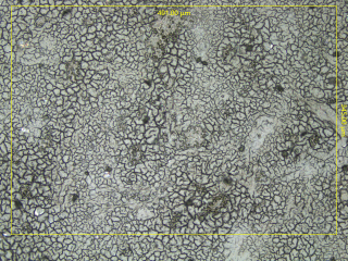
Figure 1. Microscopic image of a chicken liver cryo-section. Dark spots are suspected to be microplastic particles. Image Credit: Bruker Optics
Results
The scanned area extended to roughly 491 × 345 µm, with a spatial sampling resolution of 1.2 µm, providing a dataset able to resolve micrometer-scale inclusions present in the surrounding tissue.
An overall measurement time of 15 hours (Figure 2, top right) was planned for a traditional grid-based Raman mapping, to allow for complete coverage.
As the target particles were predicted to be scarce and irregularly dispersed, Random Scanning was used to prevent wasting measurement time on regions of the sample devoid of applicable signals.
By prioritizing the sampling of chemically detailed locations inside the chosen area, this technique increased the chances of identifying polyethylene particles while decreasing the time needed to capture informative results.
Within the first three to four hours, the software had already found and classified a large number of polyethylene particles (Figure 2, top left). The Raman image generated, even at this early stage, was very similar to the end result in both statistical validity and spatial distribution.
At every marked pixel, the typical Raman signature of polyethylene was observed. Direct evidence of polyethylene microplastics inside the liver tissue was shown in the related full-range spectra (Figure 2, bottom), which confirmed the particle’s chemical composition.
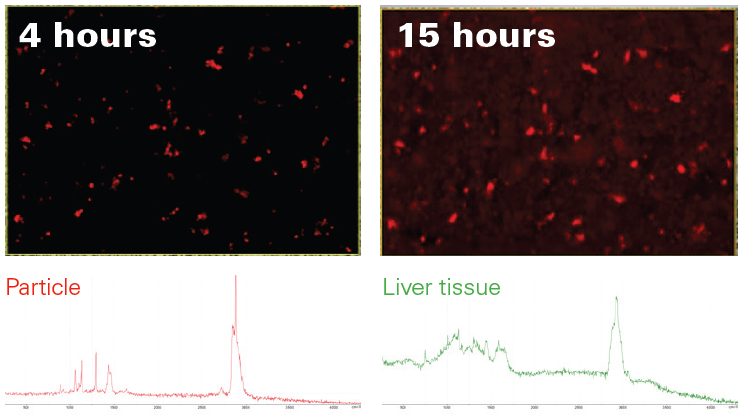
Figure 2. Comparison of the Raman image acquired with Random Scanning after four hours (top left) with the complete 15-hour Raman image (top right). Clearly, most particles have already been found by the algorithm. Found particles were unambiguously identified as PE (bottom left) and clearly stand out from the liver tissue matrix (bottom right). Image Credit: Bruker Optics
Conclusion
By coupling confocal Raman microscopy and adaptive Random Scanning, this investigation was able to deliver the direct locating and confirmation of polyethylene microplastics within liver tissue.
Raman microscopy is especially well-suited to the characterization of fine particles in complex biological structures, where high spatial resolution is fundamental. It offers an effective complementary approach for investigations that exceed the resolution thresholds of infrared techniques.
Infrared imaging continues to be a fundamental tool in the investigation of microplastics due to its unparalleled measurement speed and high-throughput screening characteristics.
Acknowledgments
Produced from materials originally authored by Dr. Yan Di from Bruker Optics GmbH & Co. KG.

This information has been sourced, reviewed, and adapted from materials provided by Bruker Optics.
For more information on this source, please visit Bruker Optics.