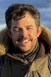
In this interview, AZoMaterials speaks with Dr. Liam Spillane, Application Scientist at Gatan, about how recent developments in counted Electron Energy Loss Spectroscopy (EELS) are transforming materials analysis.
Can you please introduce yourself and your role at Gatan?
My name is Liam Spillane - I’m an Application Scientist at Gatan, based in Pleasanton, California.
I’ve worked in electron microscopy for just over 18 years. I started my Ph.D. at Imperial College London in 2006, focusing on characterizing oxide thin films using aberration-corrected (S)TEM and monochromated EELS, and after that project and a post-doc, joined Gatan in 2012. I’ve worked with both the UK and US teams at Gatan, specializing in STEM, EELS, and the Gatan Imaging Filter (GIF) product lines.
What are the key challenges when it comes to detecting EELS signals across a broad energy range?
EELS covers an exceptionally wide energy-loss range - from millielectronvolts (meV) up to kiloelectronvolts (keV) - with the signal intensity spanning up to eight orders of magnitude. This presents a major challenge for detection.
The zero-loss peak is extremely intense, while high-energy loss edges like transitional metal K-edges are many orders of magnitude weaker. A suitable detector has to offer high energy resolution, a wide energy detection range, and extremely high dynamic range - all while minimizing noise.
How does DualEELS help address these detection challenges?
DualEELS enables near-simultaneous acquisition of low-loss and core-loss spectra using different exposure times. This effectively extends both the usable energy range at a given energy resolution as well as the dynamic range, which allows detection of strong and weak signals in the same dataset.
It has become a standard technique for many users because it provides access to critical information such as sample thickness, absolute quantification, and deconvolution. However, it comes with trade-offs - the acquisition time and total dose are increased, since two exposures are recorded instead of one.
What benefits do modern direct detection cameras like the K3 bring to EELS acquisition?
The direct detection and sharp point spread function of the K3 allow high spectral resolution and a wide energy range simultaneously in a single acquisition.
Unlike traditional fiber-coupled cameras, which lose energy resolution at low spectral magnification (which is what we mean by energy dispersion), the K3 maintains energy resolution under low dispersion conditions. This makes it possible to capture core-loss edges spanning thousands of electron volts whilst maintaining source or close to source, energy resolution - i.e., 0.4 eV with a CFEG. Multiple core loss acquisition ranges really aren't required using the K3. Capturing more information in a single readout is the fastest and most dose-efficient we can do. This is highly beneficial for all STEM-EELS experiments, but becomes absolutely critical for beam-sensitive samples. Remember - if we can collect the information in one spectrum, that’s ideal. Two is a compromise, and more than two is even more of a compromise.
How does the K3 enable dose fractionation, and why is that important?
The K3 combines fast acquisition speeds - down to 339 microseconds per frame, with near-zero noise readout - making it possible to spread the total electron dose across many short exposures. i.e., large numbers of relatively sparse or low intensity frames can be summed post-acquisition for a high SNR final dataset.
This approach, known as dose fractionation, is well established for imaging in cryo-EM. The method enables high-speed motion correction, which improves spatial resolution in the final images, but also enables data acquisition from beam-sensitive materials. The dose fractionated data can be collected at a low dose rate, which is advantageous. That data is stored in a "dose fractionated" time series, so compromised frames can be discarded post-acquisition. The same methodology followed in STEM-SI allows acquisition of high-quality, atomic-resolution data that would otherwise be difficult or impossible to obtain.
Can you share an example of a challenging experiment made possible by these technologies?
Yes, a great example is our work on strontium ruthenate (SRO) thin films grown on dysprosium scandate (DSO) in collaboration with Berit Goodge, who is currently based at the Max Planck Institute for Chemical Physics of Solids in Dresden, Germany. DSO is extremely radiation hard and easy to work with, but SRO is very beam-sensitive for a functional oxide film. To add to the challenge, SRO is a low-temperature quantum oxide, so this work was done at liquid nitrogen temperature using a side-entry liquid nitrogen holder. In spite of these two major challenges, using a K3 counting camera, dose fractionation, and fast drift correction, we were able to perform atomic-resolution EELS mapping up to 2,800 eV energy loss, with a STEM probe current of just 20 pA - something that would have failed with conventional cameras.
Why is single-range EELS acquisition advantageous over multiple spectral ranges?
Single-range acquisition is more efficient in both dose and time. It avoids the need to segment the spectrum or expose the sample multiple times, which can lead to overdosing without full signal continuity. The sharp point spread function and high pixel count of the K3 make it possible to capture broad energy loss windows - up to 3,000 eV - with high resolution in a single acquisition. This eliminates the need for multi-range setups and simplifies post-processing.
What implications do these developments have for industrial or high-throughput labs?
Speed and efficiency are critical in labs handling large sample volumes or working under tight timelines. Counted EELS and modern direct detection cameras allow high-quality data to be acquired much faster, with reduced beam damage. We've seen the benefits of a challenging liquid nitrogen temperature experiment, but the same benefits would apply to routine analysis. High-speed continuous scanning and dose fractionation mean that samples can be put in the microscope, and we start acquiring data immediately - no need to wait for long periods of time for sample stabilization.
How do you approach post-acquisition drift correction in practice?
Live drift correction is fully automated. Post-drift correction is semi-automated. If post-acquisition drift correction is required (it isn't always), I typically use the ADF signal, which has the highest signal-to-noise ratio, to measure drift, then apply the resulting drift profile to the full spectrum image.
The software provides options for image filtering and alignment settings, making the process straightforward and efficient. I have presets saved for atomic resolution mapping that are pretty robust and reliable. It’s a user-friendly workflow, and we’re planning to release a tutorial on it soon.
How can researchers access the data or methods you have discussed?
Many of the examples that we have discussed are available as experiment briefs or webinar recordings on gatan.com.
I’ll also be presenting this work at upcoming MMC and M&M conferences - these are great opportunities to view the data in detail and discuss the methods directly. We're always happy to work with users at the microscope to help them get the most out of these tools in real time.
About Dr. Liam Spillane
Dr. Liam Spillane is an Application Scientist at Gatan, where he specializes in STEM, EELS, and GIF systems. He earned his Ph.D. in materials science from Imperial College London, focusing on aberration-corrected transmission electron microscopy (TEM) and monochromated EELS of oxide thin films.
Dr. Spillane’s postdoctoral work further explored structural and chemical characterization techniques applied to diverse materials, including ferroelectrics, catalysts, and solid oxide fuel cells. He joined Gatan in 2012, initially supporting customers in the United Kingdom before relocating to California in 2018.
Now based at Gatan’s headquarters in Pleasanton, Dr. Spillane works closely with the product and software teams, contributing to advanced detector development and applications.
His research and teaching frequently focus on optimizing dose efficiency and expanding the capabilities of EELS in challenging environments such as cryogenic and in situ studies. He is a regular contributor at microscopy conferences such as M&M and MMC.

This information has been sourced, reviewed, and adapted from materials provided by Gatan, Inc.
For more information on this source, please visit Gatan, Inc.
Disclaimer: The views expressed here are those of the interviewee and do not necessarily represent the views of AZoM.com Limited (T/A) AZoNetwork, the owner and operator of this website. This disclaimer forms part of the Terms and Conditions of use of this website.