Whether in industrial fluids, fuels, biopharmaceuticals, or advanced materials, understanding particle behavior is fundamental to performance and quality.
Image-based particle analysis has become increasingly important over the past decade because it provides more information than traditional methods such as laser diffraction, light obscuration (LO), or dynamic light scattering.
Two image-based approaches are often mentioned when discussing these technologies:
- Micro-flow imaging (MFI®), which is widely used in biopharmaceutical research
- Dynamic Image Analysis (DIA), which is a broader and more scalable technology utilized in both research and high-throughput QC environments
Micro-flow imaging and flow-image microscopy are employed by a wide range of equipment manufacturers. (MFI® is a registered trademark of ProteinSimple, a Bio-Techne brand).
While both of these methods capture particle images in flow, their workflow suitability, design philosophy, and scalability are fundamentally different from DIA.
This article explains these key differences, examining why dynamic image analysis is becoming the solution of choice for laboratories seeking accurate, scalable, and production-ready sub-visible particle data.
Micro-Flow Imaging: A Research-Focused Tool Developed Around Very Low Sample Volumes
Micro-flow imaging was initially developed to support the study of protein aggregation and other sub-visible particles used in formulation development. Early-stage biopharma organizations often work with small batch sizes, so MFI systems were optimized to accommodate low particle loads and microliter-scale sample consumption.
These low sample requirements make MFI highly effective for addressing an array of research questions, including:
- Looking at how a formulation behaves under freeze–thaw stress
- Determining whether protein aggregates are reversible or irreversible
- Checking whether silicone oil droplets appear during agitation
These design choices can introduce workflow limitations for scale-up efforts, however, including quality control, manufacturing, and high-volume testing.
Real-World Research, QC, and Manufacturing Environments
Microliter sample volumes are typically incompatible with real-world sampling, with quality control and release testing often necessitating the use of representative sample volumes rather than tiny microliter aliquots.
Low sample volumes and slow flow rates can also limit:
- Homogeneous sampling of larger containers
- Scalable sampling from IV bags
- Testing from prefilled syringes
- High-volume batch screening in manufacturing
Low-throughput design can cause further issues, with low flow rates making this approach:
- Slow when working with large sample sets
- Challenging to integrate into existing release testing workflows
- Inefficient when used for in-process or continuous monitoring
Sensitivity to particle load is frequently challenging because real biopharma samples often contain:
- Surfactant micelles
- Silicone droplets
- Aggregates
- Air bubbles
- Excipient crystals
Samples may settle out or rise to the top of the sample entry port and escape analysis when low sample volume and slow flow rates are utilized.
Micro-flow imaging systems generally operate at very low flow rates to maintain image sharpness within the small optical cell, but slow flow rates also allow particles time to settle prior to imaging.
Slow flow movement creates a physical problem, whereby particles with even small density differences (for example, silicone droplets, glass shards, protein aggregates, and polymer fragments) start to settle or rise in the syringe or inlet tubing before reaching the imaging window.
There is also a risk that surface-active particles, such as silicone droplets or protein aggregates, may adhere to the tubing walls, preventing them from entering the flow cell. When this happens, the population reaching the camera may not match the actual population in the container, causing issues with representativeness.
It is also important to note that low sample volumes will not produce statistically representative sub-visible particle data. This is especially notable in real-world biopharmaceutical sampling, where particle distributions are often heterogeneous, particularly in the case of:
- Prefilled syringes
- IV bags
- Bulk drug substance containers
- Final drug product batches
Micro-flow imaging workflows typically use just microliters to a few milliliters of sample, which significantly reduces the likelihood that the drawn sample accurately represents the full container.
Small volumes increase the chances of:
- Missing rare yet critical particle types
- Overestimating or underestimating true sub-visible particle counts
- Sampling errors arising from stratification or settling
- Failing to capture low-frequency or intermittent contaminants
- Missing glass particulates from needle abrasion or stopper coring
Representative sampling is critical for quality control and release testing, but this is a key limitation of micro-flow imaging.
Dynamic Image Analysis (DIA): A Scalable, Production-Ready Alternative
Dynamic Image Analysis directly addresses micro-flow imaging’s inherent scalability and workflow limitations. Both technologies work by imaging particles while in motion, but DIA has been developed to accommodate a much wider range of sample volumes, types, viscosities, workflows, and particle concentrations.
Systems like the Raptor 1788 offer a range of advantages.
Compatibility with Both Microliter and High-Volume Samples
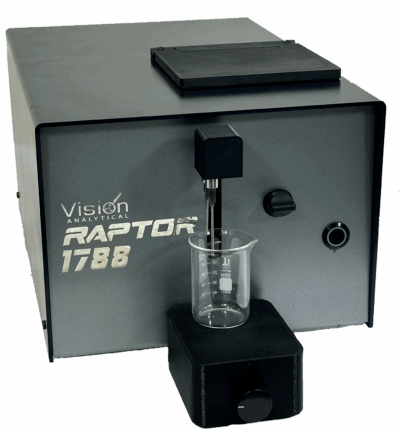
Image Credit: Vision Analytical Inc.
Whether analyzing just microliters or scaling up to hundreds of milliliters, DIA enables flexible sampling to meet diverse analytical requirements.
This flexibility allows direct measurement of:
- Prefilled syringes (for non-destructive testing)
- IV bags (for non-destructive testing)
- Bulk drug substance containers
- In-process streams
- Larger manufacturing batches
This degree of flexibility and compatibility is one of DIA’s most notable advantages over micro-flow imaging and flow-image microscopy.
Support for True ‘Real-World’ Sampling
Micro-flow imaging requires that the sample be brought to the instrument in very small quantities, but DIA systems have been specifically designed to integrate with:
- Light Obscuration particle counter sampling loops
- In-process monitoring ports
- Continuous flow systems
- Small volume syringes
- Syringe pump interfaces
- Larger industrial reservoirs
This potential for integration offers significant workflow benefits to QC and manufacturing, including:
- Fewer sampling artifacts
- Improved representativeness
- Reduced contamination risk due to fewer transfers
- Compatibility with automated sampling systems
Higher Throughput with Maintained Image Quality
Contemporary DIA systems offer consistently strong image quality, while also allowing:
- Higher particle concentrations
- Larger volumes
- Faster flow rates
- Continuous or semi-continuous sampling
These capabilities mean that DIA is suitable for:
- Stability testing
- Batch release testing
- Verification of line clearance
- Routine monitoring of contamination
Broad and Accurate Morphology Characterization
Micro-flow imaging and DIA both provide morphology data, but DIA generally offers a more comprehensive feature set, including:
- Aspect ratio
- Solidity
- Circularity
- Convexity
- Fiber length and width
- Particle type classification based on over 30 shape models
- Particle Correlation Plots are designed to detect rare events
This diverse range of shape metrics helps users to distinguish:
- Aggregates versus silicone droplets
- Glass shards versus polymer fragments
- Fibers versus elongated protein clusters
- Bubbles versus true particles
This is often the deciding factor for root-cause analysis.
Advantages of DIA
DIA is emerging as the more robust solution for sub-visible applications. This is primarily due to its:
- Improved sample representativeness, with DIA producing more statistically meaningful sub-visible particle data due to its capacity to run larger volumes.
- Suitability for QC, manufacturing, and release testing, with DIA able to adapt and scale where micro-flow imaging can be too slow.
- Ability to accommodate real-world matrices, including viscous formulations, surfactants, and high particle loads.
- Eliminate workflow bottlenecks, ensuring fewer clogs, fewer dilutions, and significantly less downtime.
- Capacity for direct sampling from production equipment.
- Improved accuracy due to higher flow rates and anti-settling motion: DIA systems’ higher flow rates prevent the sedimentation or flotation effects common in slow-flow systems.
These capabilities ensure that:
- A wider range of particles reaches the imaging zone.
- Fragile aggregates are consistently transported.
- There is a reduction in wall adhesion artifacts.
- The sample remains well-suspended and representative.
- The resulting data sets are more accurate and better reflect the true particle load.
It is also important to note that representative sample volumes reduce statistical error.
DIA can process from milliliters to hundreds of milliliters when required, ensuring the sample measured is much more representative of the actual container or batch. This significantly reduces sampling error, which is a key factor in sub-visible particle decision-making.
Reduced sampling error also offers major advantages in biologics manufacturing, where increasing value is placed on continuous inspection and in-line QC.
Conclusion
Micro-flow imaging continues to be regarded as a powerful research tool for studying protein aggregation, but its low-volume, low-throughput design means it is less suited to use in high-volume or production environments.
Dynamic Image Analysis offers all the benefits of microflow imaging while removing its limitations, allowing laboratories to:
- Analyze real-world sample volumes
- Sample directly from production systems
- Scale from R&D to QC to manufacturing
- Accommodate challenging particle loads
- Develop richer morphological insight
- Connect to Light Obscuration systems as an orthogonal method
Organizations looking for reliable, scalable, and representative sub-visible particle analysis are advised to consider implementing DIA, which is increasingly becoming the preferred and most practical analytical option.
Acknowledgments
Produced from materials originally authored by Vision Analytical Incorporated.

This information has been sourced, reviewed, and adapted from materials provided by Vision Analytical Inc.
For more information on this source, please visit Vision Analytical Inc.