Bird feathers’ coloration stems from a combination of pigmentation and microstructural optical effects functioning at spatial scales ranging from whole barbs down to individual barbules just a handful of microns across.
Characterizing these microscopic structures’ spectral reflectance necessitates the use of an instrument capable of both broadband spectral coverage and high spatial resolution.
This article details the CRAIC Technologies 2030PV PRO™ microspectrophotometer’s ultraviolet-visible-near-infrared (UV-Vis-NIR) reflectance microspectroscopy capabilities to characterize individual barbules from two feather samples exhibiting structurally distinct coloration.
Measurements were acquired at six locations on a blue feather and a bronze feather using two objectives of differing magnification. A 40x quartz objective (5.8 × 5.8 micron sampling area) was used first, followed by a 52x Schwarzschild-type reflecting objective (4.5 × 4.5 micron sampling area).
Spectra were collected over the spectral range 250–800 nm and referenced to a sintered Teflon white standard.
The two feather types were found to produce notably different reflectance profiles. For example, the blue feather showed a broad reflectance peak in the blue region of the visible spectrum that was deemed to be consistent with structural (non-pigmentary) color.
The bronze feather exhibited a reflectance profile that was found to increase monotonically with wavelength, however. This was deemed to be consistent with pigment-based absorption.
The two objectives were compared, revealing comparable spectral quality across the shared wavelength range, but noticeably improved image quality using the quartz objective at this magnification.
These results presented here highlight how aperture-limited reflectance microspectroscopy is a non-destructive and practical method for resolving the distinct optical signatures of individual feather microstructures.
Background
Bird feathers’ vivid and typically iridescent coloration has interested both biologists and materials scientists for a long time, because this coloration arises from two profoundly different physical mechanisms.
Pigmentary coloration is the result of the selective absorption of light by melanins, carotenoids, or other chemical pigments embedded within the feather structure.
Structural coloration, however, stems from the physical interaction of light with the feather’s nanoscale and microscale architectural features, for example, the keratin matrix and air-pocket arrangements within barbules.
These features produce color through scattering, interference, or diffraction rather than absorption.
Differentiating between these mechanisms and characterizing their variations across a single feather’s fine structural elements requires optical measurements at a spatial resolution far below that of the naked eye or even standard bulk spectrophotometry.
A feather is hierarchically structured: a central rachis gives rise to barbs, which in turn bear smaller barbules that are just a few microns wide.
Coloration can vary meaningfully at this barbule scale, meaning that it is necessary to use an instrument that can resolve spectral reflectance from areas of only a few square microns to accurately characterize feather optical properties at the level of individual structural units.
The study presented here showcases the use of UV-Vis-NIR reflectance microspectroscopy for this purpose, applying this powerful technique to two feather samples with visibly distinct coloration.
The spectral differences among the feathers are evaluated, and the practical performance of two candidate microscope objectives for this type of measurement is also assessed.
Materials and Methods
The 2030PV PRO™ UV-Vis-NIR microspectrophotometer (CRAIC Technologies, Inc.) (Figure 1) was used to perform reflectance measurements. This instrument was configured for reflectance spectroscopy using a permanently calibrated variable aperture system.
This configuration enables the adjustment and precise positioning of the measurement aperture over a target region of interest as small as a few microns across. Spectra can, therefore, be collected from individual barbules as opposed to from bulk feather regions that incorporate a range of structural elements.
Two feather samples were examined: one displaying blue coloration and one displaying bronze coloration.
The feathers were placed directly onto a quartz slide, with no staining, sectioning, or other preparative treatment applied. This minimal approach to sample preparation preserved the barbules’ native structure.
Measurements were conducted under incident illumination in reflectance mode, with a sintered Teflon white standard used as the reference to calculate percent reflectance.
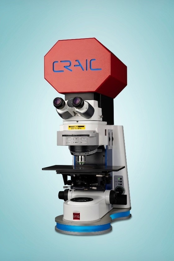
Figure 1. 2030PV PRO™ UV-Visible-NIR microspectrophotometer. Image Credit: CRAIC Technologies
Two objectives were compared to evaluate the influence of objective choice on measurement quality. A 40x quartz refractive objective was assessed, yielding a sampling area of 5.8 × 5.8 microns, and a 52x Schwarzschild-type reflecting objective was evaluated, yielding a smaller sampling area of 4.5 × 4.5 microns.
Each feather sample was analyzed with each objective at a total of six distinct barbule locations. Both reflectance spectra and co-registered microscope images were acquired at each location across a spectral range of 250 to 800 nm, spanning the near-ultraviolet through near-infrared.
Results
Representative microscope images were collated, confirming that the measurement aperture was successfully positioned on individual barbules for both the blue and bronze feather samples, for both objectives tested. The measurement aperture was visualized as a small black square in each image.
These results confirm that the achieved spatial resolution was sufficient to isolate single barbule structures from the surrounding feather architecture.
The blue feather produced a reflectance spectrum with a broad peak centered in the shorter-wavelength (blue) region of the visible spectrum when using the 40x quartz objective. Reflectance was found to decrease at both longer and shorter wavelengths prior to slightly rising again toward the red end of the range.
In contrast, the bronze feather demonstrated a reflectance spectrum that steadily rose almost monotonically from the ultraviolet through the visible range. This spectrum reached its highest reflectance values toward the longer-wavelength end of the measured spectrum. These findings are consistent with the visual appearance of a warm, longer-wavelength-dominated color.
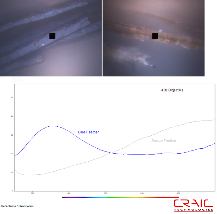
Figure 2. The reflectance microspectra were acquired from individual barbules with the 40x objective. The blue feather image is shown on the left, while the bronze feather image is on the right. Both images were acquired with incident illumination. The black square is the measurement aperture that was used for the spectroscopy. Image Credit: CRAIC Technologies
These two reflectance profiles’ general shapes were preserved when the measurement was repeated with the 52x Schwarzschild objective. This shows that the observed spectral characteristics are a genuine property of the barbule structures as opposed to an artifact of the particular objective employed.
For both samples, the overall reflectance magnitude recorded using the 52x objective was found to be lower than the reflectance recorded with the 40x objective. This is consistent with differences in aperture size and optical throughput between the two objective designs, rather than any alteration in the samples’ intrinsic reflectance.
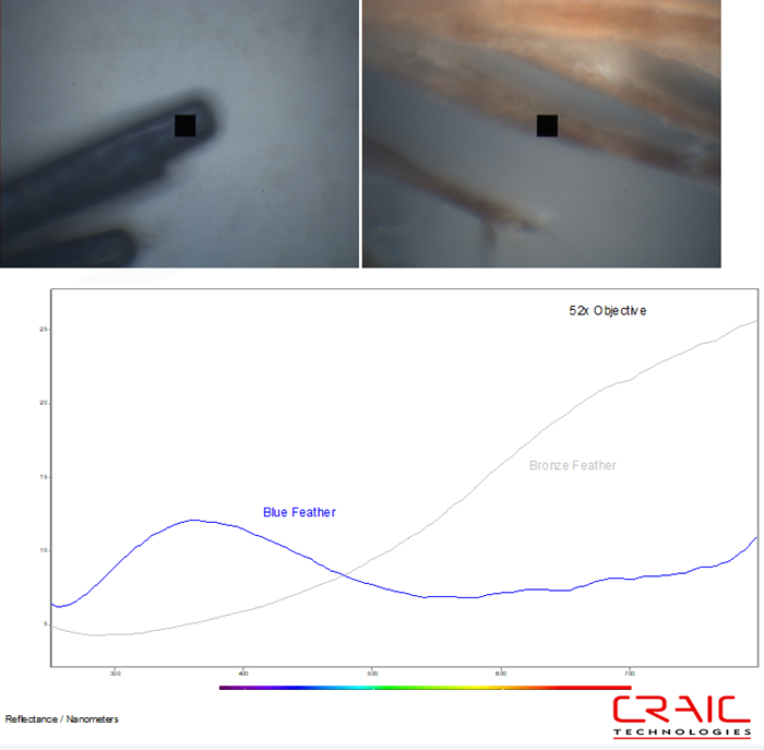
Figure 3. The reflectance microspectra were acquired from individual barbules with the 52x objective. The blue feather image is shown on the left, while the bronze feather image is on the right. Both images were acquired with incident illumination. The black square is the measurement aperture that was used for the spectroscopy. Image Credit: CRAIC Technologies
Discussion
The blue and bronze feathers’ contrasting reflectance profiles are consistent with the two coloration mechanisms described.
It was observed that the blue feather's peaked reflectance spectrum was concentrated in a relatively narrow band of shorter wavelengths. This signature is anticipated when observing structural coloration, in which light of a limited wavelength range is preferentially reflected or scattered by the barbule’s physical microarchitecture rather than by chemical absorption.
In contrast, the bronze feather's smoothly rising reflectance profile toward longer wavelengths is more consistent with pigment-based coloration, in which shorter wavelengths are progressively absorbed while longer wavelengths are reflected.
Comparing the 40x quartz and 52x Schwarzschild objectives offered practical guidance for choosing appropriate instrumentation in future barbule-level studies.
The two objectives yielded fairly similar spectral shapes across their shared spectral range, but it was noted that the image quality achieved with the quartz objective was notably superior at this magnification.
This improved image quality is a key consideration when there is a need to precisely visually target specific microstructures as opposed to a spectral range alone.
It was also observed that the Schwarzschild objective's reflective (rather than refractive) optical design offers the benefit of chromatic-aberration-free performance across a wider spectral range that extends further into the ultraviolet.
These capabilities may be preferable for studies emphasizing deep-UV and far-NIR structural features that are not well resolved in the dataset presented here.
These findings highlight the technical feasibility and biological information available using reflectance microspectroscopy at apertures of only a few square microns. This technology can be used to directly link feather microstructure with optical function, without the need to implement destructive sample preparation.
Conclusion
Performing UV-Vis-NIR reflectance microspectroscopy with a calibrated variable-aperture instrument able to resolve areas as small as 4.5 × 4.5 microns has been shown to successfully distinguish the spectral signatures of individual barbules from two feathers featuring distinct coloration mechanisms.
It was observed that the blue feather exhibited a reflectance profile that is consistent with structural color, while the bronze feather exhibited a profile that is consistent with pigment-based absorption.
Comparing two candidate objectives revealed comparable spectral performance, but the quartz objective was shown to offer improved image quality at the tested magnification.
These results reinforce the benefits of leveraging aperture-limited microspectroscopy as a non-destructive tool for investigating feather coloration’s optical basis at the scale of individual structural elements.

This information has been sourced, reviewed, and adapted from materials provided by CRAIC Technologies.
For more information on this source, please visit CRAIC Technologies.