The newest 3D X-Ray microscope models in the ZEISS Xradia Versa series offer users increased options for their scientific and industrial investigations.
The ZEISS Xradia 610 and 620 Versa push the limits of non-destructive sub-micron scale imaging by utilizing industry-leading resolution and contrast.
Highlights
Extending the Limits of Micro- and Nano-CT Solutions
- Improved flow and speedier scans without sacrificing clarity
- Non-destructive submicron microscopy of intact samples
- Image quality with high throughput
- Adaptable and scalable to future developments and additions
- The smallest possible voxel size is 40 nm, with a true spatial resolution of 500 nm.
- In situ imaging for continuous, controlled, non-destructive microstructure categorization.
- Excellent resolution across a wide range of operational ranges, proportions, and sample types
Highest Resolution and Flux
The Xradia Versa employs novel two-stage magnification optics and a high flux X-Ray source to produce images with sub-micron scale resolution faster than standard tomography, which depends on single-stage geometric magnification.
Larger, denser objects, such as full machinery and component assemblies, can be imaged in 3D with high resolution, thanks to the Resolution at a Distance (RaaD) architecture. Navigating to necessary sequences and quickly scanning big samples (up to 25 kg) are made possible by the extra flat panel extension (FPX).
New Degrees of Freedom
For advanced scientific and industrial research, use the most comprehensive 3D X-Ray imaging solution to achieve breakthrough material and property characterization, increase absorption, and improve phase contrast.
Contrast Tomography offers insights into diffraction 3D crystallographic information. Users can enhance scanning efficiency and accuracy for large or sporadic samples through advanced acquisition techniques. Additionally, machine learning methods can assist in sample post-processing and segmentation.
Premier 4D/In Situ Solution
The ZEISS Xradia 600 Series Versa can examine formation development over time (4D) and characterize 3D microstructures of materials non-destructively (in situ).
The Xradia Versa can accommodate samples, environmental chambers, and high-accuracy in situ load rigs without compromising resolution. It employs Resolution at a Distance to maintain the best resolution across extended distances.
The Versa readily integrates with other ZEISS microscopes to solve multi-scale correlative imaging problems.
Application Examples
The Versa 610 and 620 ZEISS Xradia at Work
Electronics and Semiconductor Packaging
Typical Tasks and Applications
- Examine printed circuit boards for hardware security and reverse engineering.
- Non-destructively image from module to package to interconnect for submicron defect analysis at speeds that can complement physical cross-sectioning throughout a range of length scales.
- Analyze the structure and failure of advanced semiconductor packages for process development, yield enhancement, and construction analysis. This includes 2.5/3D and fan-out packages.
- Gain a better understanding of defect locations and distributions by viewing infinite virtual cross-sections and plan view images from any desired angle.
Lithium-Ion Batteries
Typical Tasks and Applications
- Assessment for safety and quality: Identifying debris, particle formation, burrs at electrical contacts, or damage to the polymer separator.
- Impacts of aging and lifetime: Longitudinal studies on the effects of aging
- Recipe creation and supply chain management: Evaluation of complete samples for successful supplier management, exposing recipe changes or cost reductions that can reduce longevity or efficacy.
Additive Manufacturing
Typical Tasks and Applications
- Examining the internal components' surface roughness that cannot be reached by other means.
- 3D image to contrast it with the official CAD model.
- Vapors, high-Z inclusions, and unmelted particles are found.
- An extensive shape, size, and volume distribution examination of the particles in a powder bed used in additive manufacturing (AM) is carried out to determine the best process parameters.
- Microstructural analysis of AM components by non-destructive, high-resolution imaging
Materials Research
Typical Tasks and Applications
- Observe characteristics at different scales of length
- Demonstrate the three-dimensional structure
- Measure microstructural evolution.
- Examine the methods of failure, instances of degradation, and internal flaws.
- To comprehend the effects of heating, desiccation, chilling, wetting, compression, tension, drainage, imbibition, and other simulated environmental study, conduct in situ and 4D time-dependent investigations.
Raw Materials
Typical Tasks and Applications
- Users can examine the grain alignments of steel and other metals
- Particle analysis combined with full 3D reconstruction
- Analyze fluid flow and pore structure at various scales
- Examine crystal forms with LabDCT Pro
- Openly monitor fluid flow at the pore scale with in situ flow technologies
- Improve mining procedures by conducting thermodynamic leaching studies, QA/QC analysis on mining products, such as iron ore pellets, and tailings studies to optimize mining operations
Life Sciences
Typical Tasks and Applications
- 3D imaging of biological samples in their natural environment
- Imaging of delicate plants and animals without sample processing or sectioning
- Sub-micron photography of whole solid objects, such as seeds
- Imaging plant roots still rooted in their original soil without any special sample preparation
Technology Insights
Non-destructive imaging with the highest contrast and resolution possible.
Highest Resolution Without Compromise
The geometric structure of magnification in standard X-Ray computed tomography (CT) limits imaging at high resolution to small sample sizes. Long working distances make it challenging to maintain high resolution for larger samples.
Maintaining excellent resolution for large samples is challenging due to the longer working distances needed. The majority of CT manufacturers promise a maximum resolution, however, its practical application is limited.
The Xradia 600 Series from ZEISS Versa addresses these trade-offs, which combines high flux X-Ray source technology with dual-stage magnification.
ZEISS specifies a true spatial resolution to provide a single measure of microscope effectiveness for 3D X-Ray measurement. The smallest difference between two features that an imaging system can rectify is referred to as spatial resolution. Systems from the ZEISS Xradia 600 Series Versa may reach a 500 nm spatial resolution with voxels as small as 40 nm.
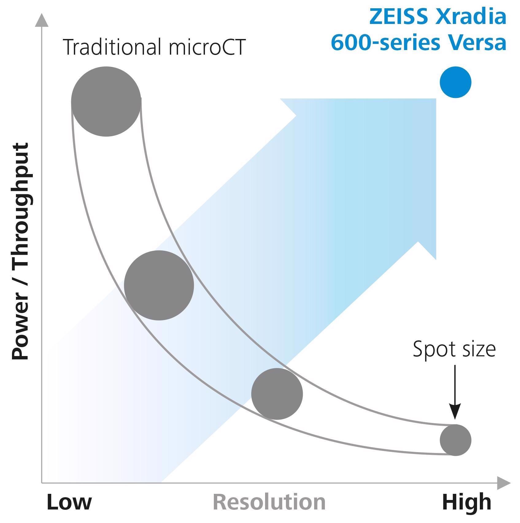
Image Credit: Carl Zeiss Microscopy GmbH
Higher X-Ray Flux Source
Numerous Advantages
With its innovative high-power (25 W) X-Ray source technology, the ZEISS Xradia 600 Series Versa substantially increases X-Ray flux over its predecessors.
The potential source pushes the envelope of efficacy with enhanced flux and throughput, resolution efficiency, and thermal management. Improved source responsiveness leads to quicker scan configuration and a more enjoyable user experience, thanks to a new source management system.
What an advanced X-Ray flux provides:
- Several sample runs
- More pronounced diffraction patterns
- Workflows for long or multiple scans (in situ, DSCoVer, stitching, DCT) are now feasible.
- Faster tomography scans
- Improved contrast-to-noise ratio
- Large areas of interest
ZEISS X-Ray Microscopes
The Versatile Advantage of RaaD
With its two-stage magnification design, the ZEISS Xradia Versa offers a wide range of sample sizes and types of sub-micron resolution imaging at long working distances or Resolution at a Distance.
As with traditional microCT, images are first geometrically magnified before being placed onto a scintillator to convert X-Rays into visible range images. These images are then optically magnified with microscope optics and captured by a CCD detector.
The ZEISS Xradia 600 Series Versa offers speedy findings for more sample sizes and types without sacrificing resolution because of its increased capacity for X-Ray photons.
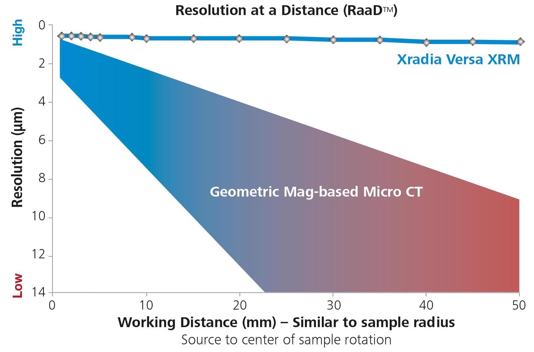
Image Credit: Carl Zeiss Microscopy GmbH
Conventional Micro CT Architecture
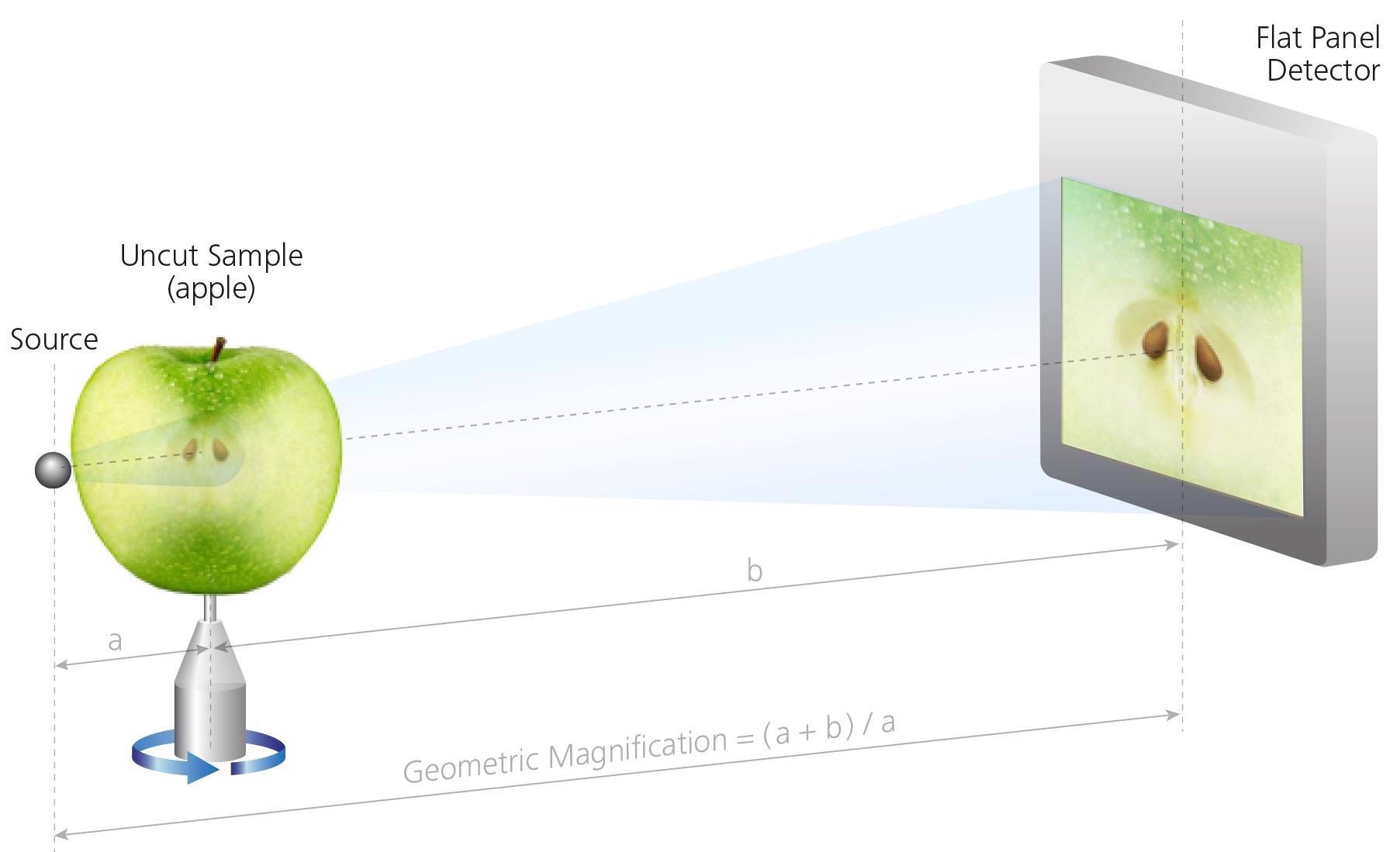
The sample must be close to the source to achieve resolution. Image Credit: Carl Zeiss Microscopy GmbH
ZEISS XRM Two-Stage Magnification Architecture
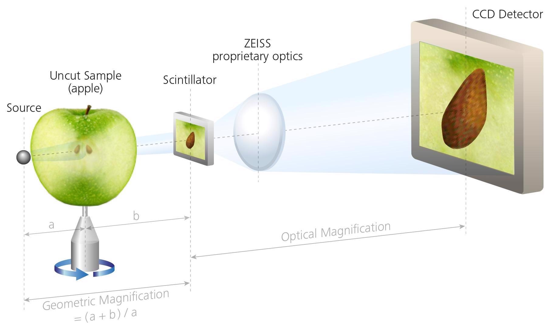
Sample imaged independently of distance to the source, enabling interiors of larger samples to be imaged non-destructively at higher resolution. Image Credit: Carl Zeiss Microscopy GmbH
Accessories
Increase the number of options available for 3D advanced material characterization.
ZEISS Advanced Reconstruction Toolbox
Increased throughput and better image quality.

Image Credit: Carl Zeiss Microscopy GmbH
Advanced Reconstruction Toolbox (ART) introduces Artificial Intelligence (AI)-driven reconstruction methods on the ZEISS Xradia 3D X-Ray microscope (XRM) or microCT. By gaining a deeper comprehension of X-Ray physics and applications, users can tackle even the most challenging imaging challenges in fresh and creative ways.
Users can investigate how ART's proprietary modules, PhaseEvolve and their versions, OptiRecon and DeepRecon, improve data gathering, reconstruction speed, and image quality without compromising resolution.
The user can employ the Advanced Reconstruction Toolbox to:
- Enhance the contrast-to-noise ratio to reveal tiny differences
- Attain improved throughput or internal tomography on a variety of samples.
- Obtain an order of magnitude increase in sample class speed that requires repetitive workflow.
- Improve data collection and analysis to enable quicker and more accurate decision-making.
- Boost the quality of the image
The optional modules are workstation-based solutions for easy accessibility and usability:
- PhaseEvolve to enhance contrast
- Iterative reconstruction with OptiRecon
- DeepRecon Pro & Custom for reconstruction based on deep learning
ZEISS DeepRecon Pro provides a straightforward, uncomplicated, and powerful application of AI and deep neural network technology for enhancing X-Ray tomography results without prior knowledge of deep learning technology. It helps us to reduce the scan time required for in situ fluid-rock interaction experiments when we need to work with long exposure times.
Dr. Markus Ohl, X-Ray Microscopy, EPOS-NL MINT, Utrecht University
LabDCT Pro
Unlocking Crystallographic Information
Diffraction contrast tomography (DCT) with LabDCT Pro allows for non-destructive 3D imaging of grain alignment and microstructure; this feature is exclusive to the Xradia 620 Versa.
The precise visualization of three-dimensional crystallographic grain orientation enhances the characterization of polycrystalline materials, such as metal alloys, geomaterials, ceramics, and pharmaceuticals.
- Combine modalities to understand structure-property interactions.
- Using 4D imaging experiments to investigate microstructural evolution
- Integrate 3D microstructural data with 3D crystallographic data.
- Specimens having crystal structures ranging from lower symmetry systems, like monoclinic materials, to cubic symmetry can be used with LabDCT Pro.
- Obtain comprehensive 3D microstructure analysis using various sample forms and larger representative volumes.
- Gather high-resolution crystallographic data using the specialized 4X DCT objective.
- Utilize the Flat Panel Extension to increase throughput and big area mapping for even larger samples (FPX)
Flat Panel Extension
With High Throughput, Image Significantly Larger Samples
The greatest image quality possible from ZEISS can be achieved with large-sample, high-throughput scanning with the optional Flat Panel Extension (FPX). With a feature-rich solution for business and academic research, FPX improves workflow efficiency and image adaptability.
Scout-and-Zoom is a ZEISS X-Ray microscope capability that leverages FPX to conduct exploratory "Scout" scans across a wider range, enabling the identification of interior objects of interest for subsequent higher resolution "Zoom" scans, all without the requirement for intricate sample processing.
The LabDCT Pro's high throughput and broad area mapping capabilities are also available with FPX.
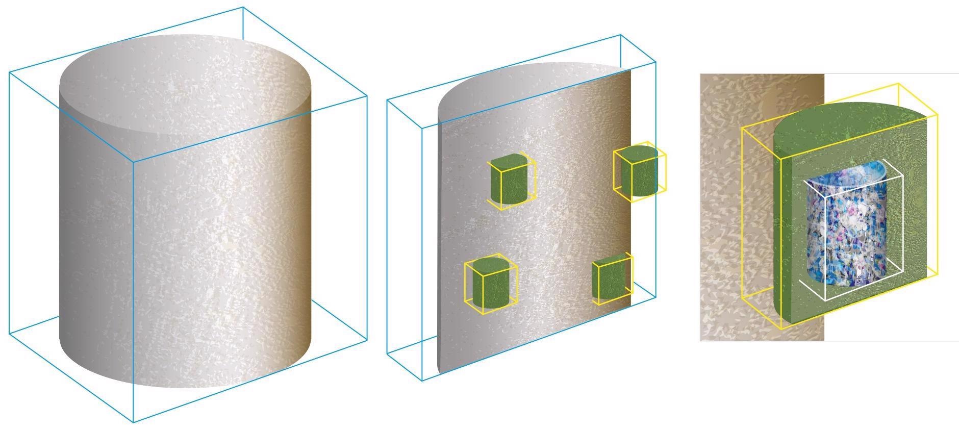
Three-stage Scout-and-Zoom workflow. Image Credit: Carl Zeiss Microscopy GmbH
in situ Experiments
Push the Limits for Scientific Development
The ZEISS Xradia X-Ray system is an industry-leading 3D imaging solution tailored for various in situ rigs, including high-pressure flow cells, strain, pressure, and heat stages.
Expanding research beyond the traditional three dimensions of space, 4D experiments leverage the non-destructive capabilities of X-Ray analysis to extend investigations into the dimension of time. The ZEISS Xradia XRM platforms offer a diverse array of in situ rigs, including highly pressurized flow cells, tension, compression, and temperature stages, as well as customizable designs to accommodate user-specific requirements.
The ZEISS Xradia XRM can be enhanced with an optional in situ Interface Kit, featuring a robotic integration package, comprehensive wiring guidance, additional facilities such as feed-throughs, and recipe-based software for seamless operation directly within the Scout-and-Scan user interface.
Upgrade the ZEISS Xradia XRM to an Xradia 620 Versa X-Ray microscope to harness the advantages of Resolution at a Distance (RaaD) technology. This innovation maximizes efficiency in tomographic imaging of samples within in situ chambers or rigs, particularly when the resolution limits of in situ studies need to be extended.
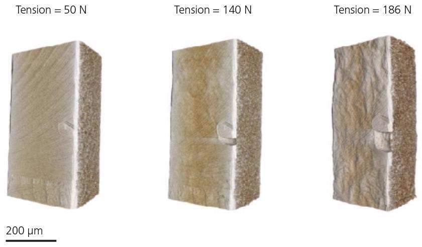
Tensile testing of laser welded steel under increasing load. Image Credit: Carl Zeiss Microscopy GmbH
SmartShield
Protect the Sample Easily to Optimize Experiment Setup
SmartShield is a comprehensive solution that safeguards both the sample and the microscope. This collision avoidance system operates seamlessly alongside the Scout and Scan Control System, empowering users with enhanced management capabilities for Xradia Versa. By simply pressing a button, SmartShield generates a digital protective coating tailored to the dimensions of the sample, ensuring greater confidence in operation.
SmartShield offers the following benefits:
- Superior scan clarity
- Safeguarding important samples and investments
- Better operator efficacy is made possible by an easy sample setup
- Improved usability for both inexperienced and seasoned users
Metrology Extension
Increasing the Accuracy of Measurement in X-Ray Microscopy
The Metrology Extension (MTX) seamlessly transforms the Xradia 620 Versa into a certified measurement accuracy solution, exceeding the limitations of standard CT technology. This is essential for industrial and academic laboratories, where the trend toward component downsizing and integration is fueling the demand for high-resolution metrology. Utilizing high-resolution X-Ray imaging alongside precise metrology provides a superior solution.
XRM Check: ZEISS has developed a (multi-sphere) length standard for verifying the accuracy of the CT measurements of small-scale dimensions. Image Credit: Carl Zeiss Microscopy GmbH
Reveal Smallest Dimensions
Measure Them Accurately
- After calibration, the user takes accurate measurements and provides the data to a standard metrology program for additional processing.
- Calibration workflow is simple: An integrated workflow for user-guided calibration is part of the MTX package.
- Compact volumes, high resolution: The MTX facilitates high-dimensional accuracy measurements within reconstructed volumes of up to 125 mm³.
- Leading CT metrology precision: The ZEISS Xradia Versa, calibrated with MTX, offers a maximum permissible error value for measurements in small-scale volumes that are competitive in the market: MPESD = (1.9 + L/100) μm, where L represents the measured length in mm.
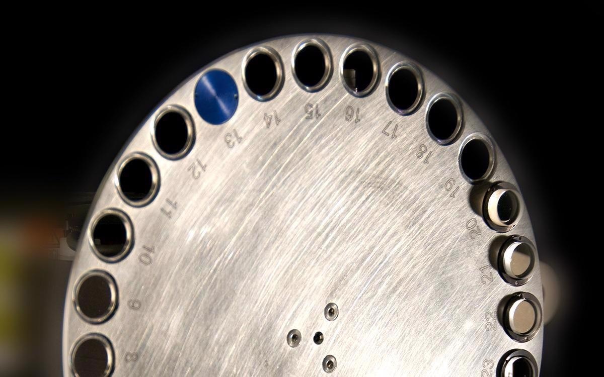
Autoloader
Improve the Efficiency of Sample Handling
Enhance the instrument's usability by incorporating the optional Autoloader, which is compatible with all ZEISS Xradia X-Ray microscope platforms.
By queuing many jobs at once, users can increase efficiency while reducing the frequency of engagement. Up to 14 sample stations, each holding 70 samples, can be loaded and set to run for a few days or overnight.
High-volume quantitative recurrent scanning of identical samples is made possible by mechanical stability that has never been seen in the industry before.
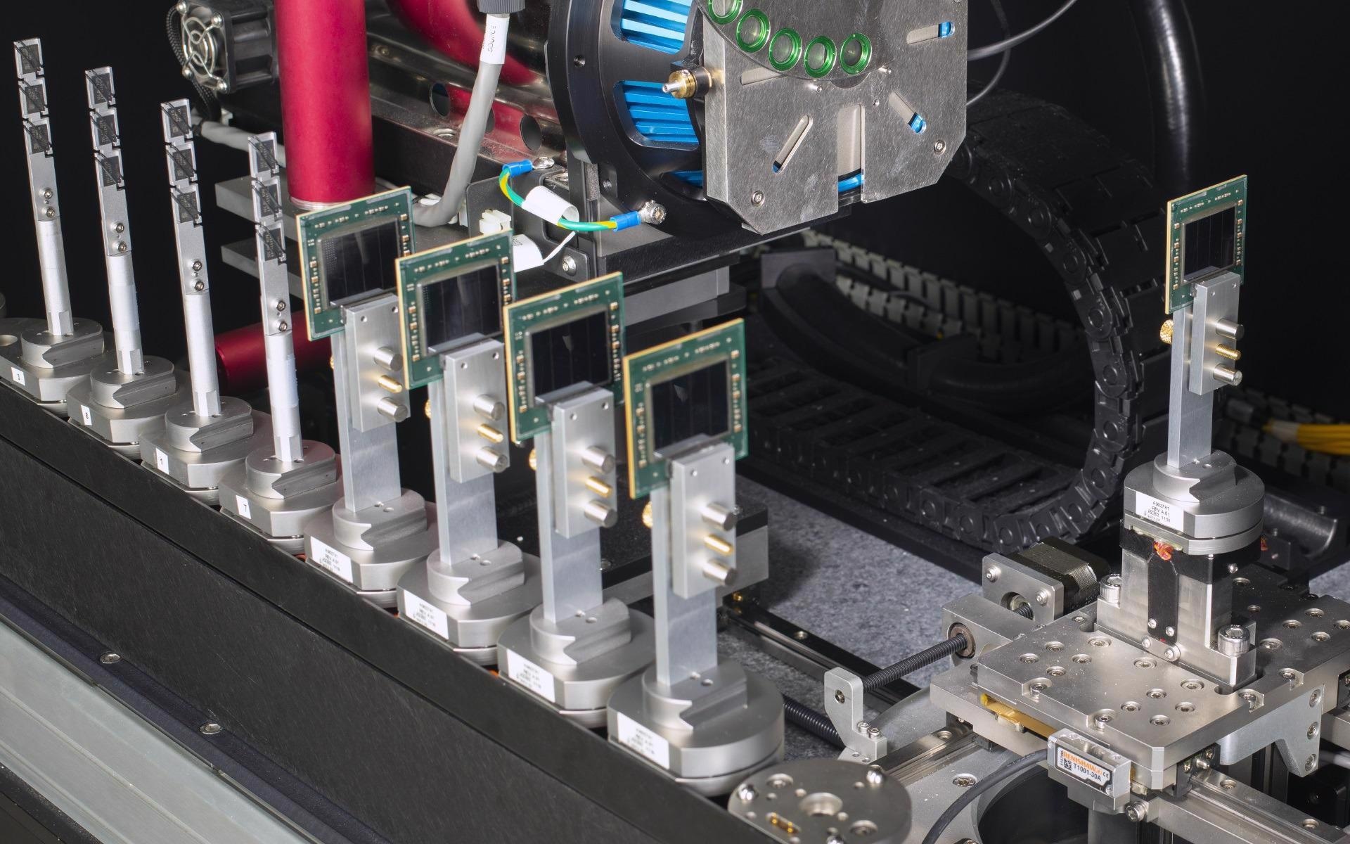
The autoloader option enables the programming of up to 70 samples at a time to run sequentially. Image Credit: Carl Zeiss Microscopy GmbH
Wide Field Mode
Image Larger Samples with Greater Flexibility
Wide Field Mode is required for users to take pictures with a strong lateral visual field (WFM). For big samples, the wide lateral field of view can offer a 3× larger 3D volume or a higher voxel density than a basic field of view.
All Xradia Versa systems feature Wide Field Microscopy (WFM) with the 0.4× objective. Additionally, the Xradia 620 Versa system offers WFM with a 4× objective. When paired with Vertical Stitching, WFM empowers users to capture high-quality images of larger samples.
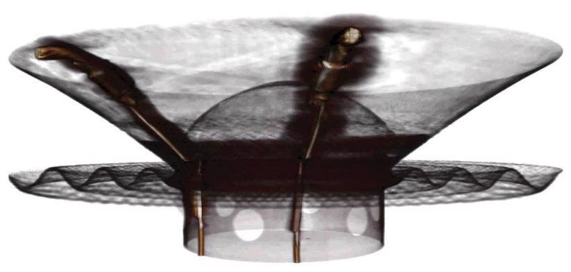
Image large samples with Wide Field Mode like this 6” stereo speaker. Image Credit: Carl Zeiss Microscopy GmbH
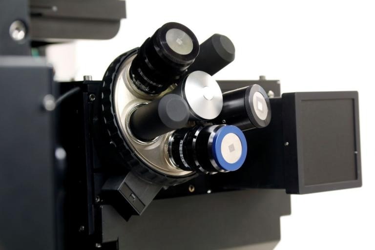
Automated Filter Changer
Simplify the Examination of Difficult Samples
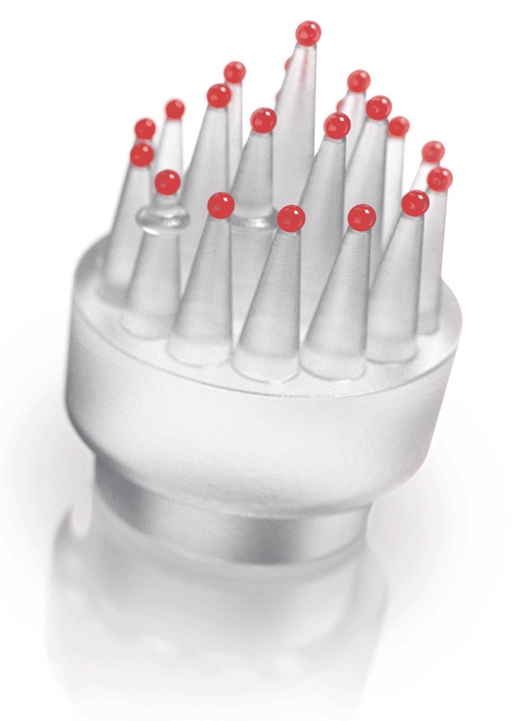
To enhance contrast, X-Ray source attenuation filters are employed to adjust the X-Ray energy spectrum, emphasizing the sample's characteristics, which depend on its unique material properties. Each ZEISS Xradia Versa is equipped with a standard set of 12 filters.
The ZEISS Xradia 610 Versa features a single filter slot for manual shifting. Meanwhile, the ZEISS Xradia 620 Versa systems incorporate an Automated Filter Changer (AFC), ensuring effortless filter adjustments to facilitate the investigation of unknown materials.
The Automated Filter Changer (AFC) offers 12 standard filters with room for 12 custom filters. Image Credit: Carl Zeiss Microscopy GmbH
The Xradia 600 Series Versa
Source: Carl Zeiss Microscopy GmbH
| |
ZEISS Xradia
610 Versa |
ZEISS Xradia
620 Versa |
| Spatial resolutiona |
500 nm |
500 nm |
Resolution at a Distance (RaaD™)a,b
(at 50 mm working distance) |
1.0 μm |
1.0 μm |
Minimum Achievable Voxelc
(Voxel size at sample at maximum magnification) |
40 nm |
40 nm |
| Source Voltage Range |
30–160 kV |
30–160 kV |
| Source Maximum Power Output |
25 W |
25 W |
| Scout-and-Scan™ Control System |
✓ |
✓ |
| Scout-and-Zoom |
✓ |
✓ |
| ZEISS OptiRecon |
Optional |
Optional |
| ZEISS DeepRecon |
Optional |
Optional |
| SmartShield |
✓ |
✓ |
| Vertical Stitch |
✓ |
✓ |
| XRM Python API |
✓ |
✓ |
| Automated Filter Changer (AFC) |
|
✓ |
| High Aspect Ratio Tomography (HART) |
|
✓ |
| Dual Scan Contrast Visualizer (DSCoVer) |
|
✓ |
| Wide Field Mode |
0.4x |
0.4x and 4x |
| ZEISS LabDCT Pro for Diffraction Contrast Tomography |
|
Optional |
| ZEISS Autoloader |
Optional |
Optional |
| In Situ Interface Kit |
Optional |
Optional |
| ZEISS ZEN Intellesis |
Optional |
Optional |
| ORS Dragonfly Pro |
Optional |
Optional |
| ZEISS Metrology Extension (MTX) |
|
Optional |
aZEISS Xradia 2D resolution target, normal field mode, and optional 40× objective were used to test spatial resolution.
b The RaaDTM working distance is defined as the clearance around the rotating axis.
c Voxel is a geometric word that adds to resolution but does not determine it, and is used solely for comparative purposes.
The genuine overall measurement of instrument resolution, as defined by ZEISS, is spatial resolution.
Protect the Investment
To safeguard customers' investments, ZEISS X-Ray microscopes are designed to be adaptable and expandable with future innovations and breakthroughs. This ensures that advancements in cutting-edge technology will be reflected in the microscope's capabilities.
Customers can field-convert their systems to the newest X-Ray microscopes with ZEISS Xradia Context microCT, ZEISS CrystalCT, ZEISS Xradia 510/520 Versa, and now ZEISS Xradia 610/620 Versa.
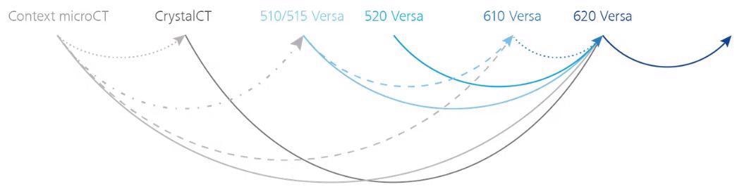
Image Credit: Carl Zeiss Microscopy GmbH
Software
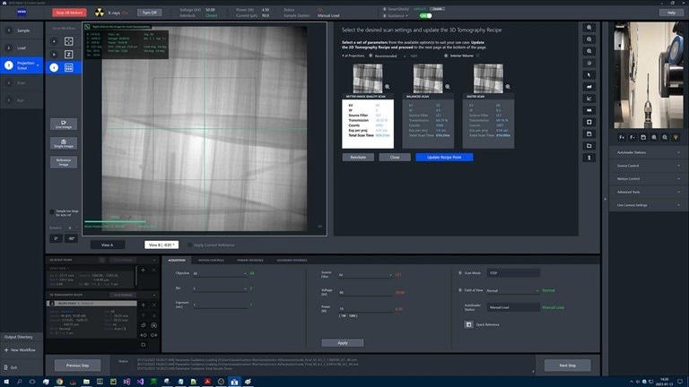
Use the Simple Control System to Create Efficient Workflows
The Scout-and-Scan Control System enables users to efficiently scout a target area and define scanning parameters. This user-friendly system is ideal for deployment in central labs, accommodating users with varying levels of expertise.
Users can benefit from the following:
- It is possible to microposition with just one mouse click
- Autoloader option for several samples
- Sample viewing using an internal camera
- Recipe management (set, save, recall)
- Different energies
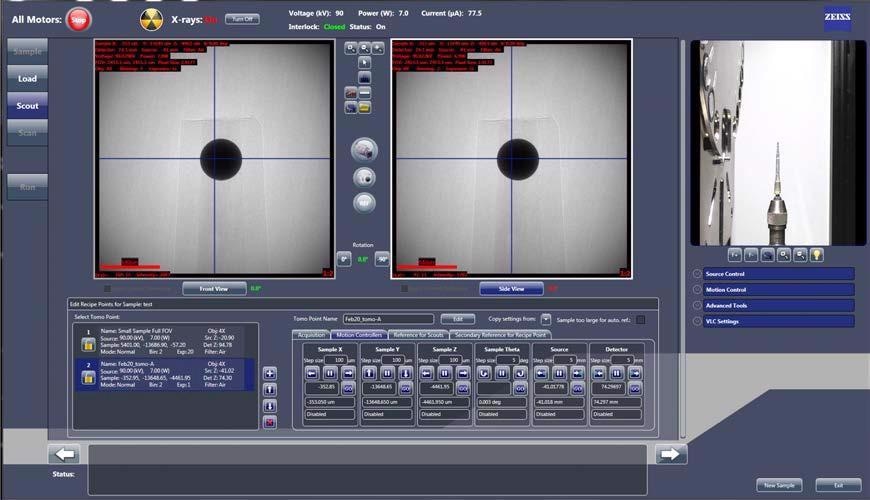
Scout-and-Scan Control System. Image Credit: Carl Zeiss Microscopy GmbH
ZEISS Mineralogic — Automated Mineralogy
ZEISS SEM, XRM, and microCT devices can be used for phase identification and textural analysis in 2D and 3D.
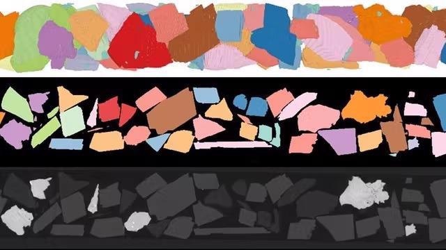
Image Credit: Carl Zeiss Microscopy GmbH
Visualization and Analysis Software
ZEISS suggests using the state-of-the-art analysis and visualization software called Dragonfly Pro from Object Research Systems (ORS) for 3D data acquired from several technologies like X-Ray, SEM, FIB-SEM, and helium ion microscopy.
Large-scale 3D grayscale data analysis and visualization are made easy with the extensive, adaptable, and user-friendly ORS Dragonfly Pro toolbox only offered by ZEISS. It enables video production, annotation, navigation, and the creation of media files from 3D data. Segmentation, object analysis, and image processing can all be used to assess the outcomes.
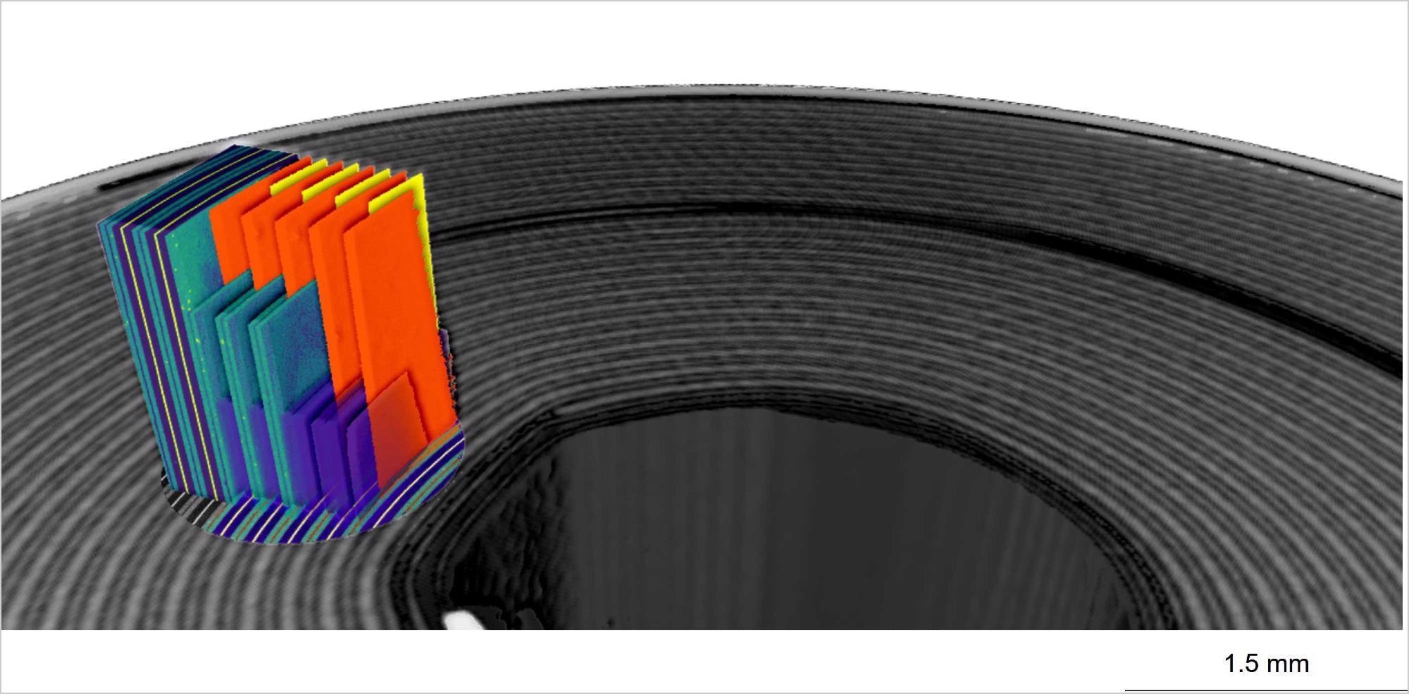
Image Credit: Carl Zeiss Microscopy GmbH
Xradia 630 Versa
With the 40X Prime objective's special higher energy capabilities, the ZEISS Xradia 630 Versa lets users achieve submicron imaging at a level never possible before. The system opens up new study possibilities with its unmatched 450–500 nm resolution performance over the whole energy range of 30 kV to 160 kV.
To deliver results quickly and effectively, NavX uses intelligent system insights to guide users via automated workflows. DeepScout, an AI-based solution, revolutionizes sample interpretation by boosting throughput 100 times faster.
 View the Versa 630 Product Brochure Here
Breakthrough Resolution Performance to Expand Research Horizons
The ZEISS 40X-Prime Objective
With increased X-Ray photon availability on the ZEISS Xradia 600-series Versa, achieving faster time-to-results for diverse samples is now possible without sacrificing resolution. Exclusive to the ZEISS Xradia 630 Versa is the 40X-Prime (40X-P) objective lens.
The ZEISS Xradia 630 Versa XRM, featuring the enhanced energy capabilities of the exclusive 40X-Prime (40X-P) objective, empowers users to push the boundaries of submicron imaging to unprecedented levels. Renowned for their Resolution at a Distance (RaaD™) capability, ZEISS Xradia Versa platforms facilitate high-resolution imaging of various sample types and sizes across an extensive range of length scales.
RaaD 2.0 is defined by the system's unmatched resolution performance of 450–500 nm with 40X-P over the whole source voltage range of 30 kV to 160 kV. The ZEISS 40X-P objective pushes industry standards for submicron imaging resolution with the ZEISS Xradia 630 Versa, opening up new application options for researchers.
Head of objectives with the 40X-P. Image Credit: Carl Zeiss Microscopy GmbH
NavX User Interface
Understanding the complexities of X-ray imaging, ZEISS XRM researchers delved into user habits and challenges, applying human-centered design (HCD) principles. The result, NavX™, the new user interface for ZEISS Xradia 630 Versa, facilitates immediate productivity even for novice users in busy environments.
NavX™ guides users through automated workflows with intelligent system insights, streamlining the delivery of experimental results while offering experienced users the flexibility to explore the platform's full capabilities.
With NavX, users can automate workflow and receive information on how the settings selected will affect the setup. The program incorporates this information immediately, guiding decisions comfortably and intuitively.
The NavX File Transfer Utility (FTU) automatically transfers microscope-generated data to other locations, ensuring users can access their data wherever and whenever needed. These enhancements significantly enhance NavX's capabilities for remote operation, thereby boosting user productivity.
NavX offers intuitive navigation, evolving alongside the XRM user base to revolutionize X-Ray navigation and control. It seamlessly integrates workflows, enhancing the planning and execution of advanced correlative workflows.
NavX User Interface. Image Credit: Carl Zeiss Microscopy GmbH
Flat Panel Extension
The ZEISS Xradia 630 Versa X-Ray microscope has a flat panel extension (FPX) as standard equipment. This adds to the system's versatility by directly supporting DeepScout, the AI-based tool for deep learning and neural network training in the Advanced Reconstruction Toolbox.
Use FPX to identify interior regions for better resolution "zoom" scans and perform low resolution, big field of view "scout" scans on various sample types. The Volume Scout workflow within NavX streamlines this procedure.
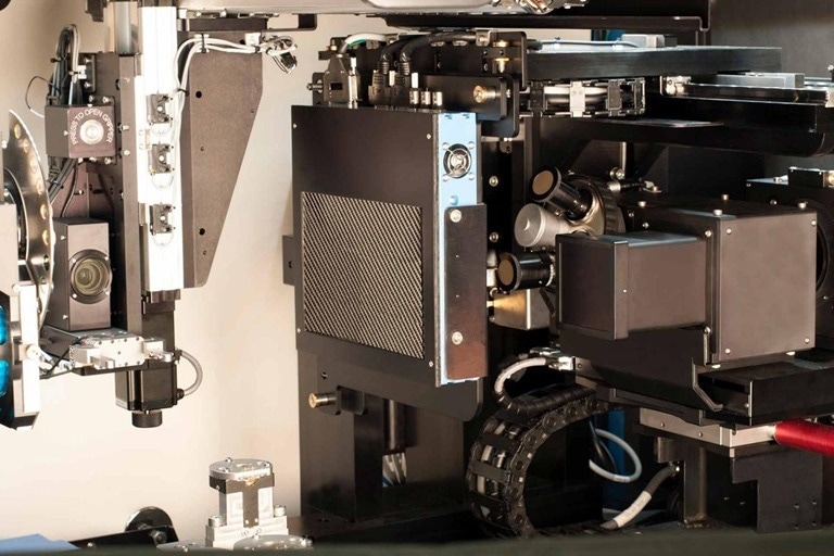
Image Credit: Carl Zeiss Microscopy GmbH
LabDCT Pro for Diffraction Contrast Tomography (DCT)
Unlocking Crystallographic Information
With Xradia 630 and 620 Versa only, LabDCT Pro for diffraction contrast tomography (DCT) allows non-destructive 3D imaging of grain orientation and microstructure. A new level in the characterization of polycrystalline materials such as metal alloys, geomaterials, ceramics, or pharmaceuticals can be unlocked by directly observing 3D crystallographic grain orientation.
- Specimens with crystal structures ranging from cubic symmetry to systems with reduced symmetry, including monoclinic materials, are supported by LabDCT Pro.
- Obtain crystallographic data with great resolution by utilizing the specialized 4X DCT objective. Extensive area mapping and the Flat Panel Extension (FPX) boost throughput for larger samples.
- Obtain thorough 3D microstructure analysis using a variety of sample geometries and larger representative volumes.
- Use 4D imaging techniques to study the evolution of microstructural features.
- Integrate 3D microstructural characteristics with 3D crystallographic data.
- Integrate modalities to comprehend links between structure and property.