ZEISS ZEN Connect software enables the integration of multiple perspectives of samples across imaging modalities and scales. Images can be organized and overlayed by the culmination of all imaging technologies. Some of the prime features of ZEN Connect are:
- Shift to confocal, light or electron microscope and align one time
- Obtain an overall image
- Overlay and align images from any source
- Save multi-modal data in well-ordered projects
- Utilize overview image for navigation
ZEISS ZEN Connect is time-efficient, offers distinctive insights and is an efficient tool for a multi-user facility — for a materials researcher at a university or an industrial laboratory.
More than one microscopy modality or contrasting techniques can be used with Zen Connect, which helps in complex applications, such as experimenting with additive manufactured parts, examining batteries for e-mobility, studying raw materials like oil or even assessing inclusions in metallic samples.
Highlights
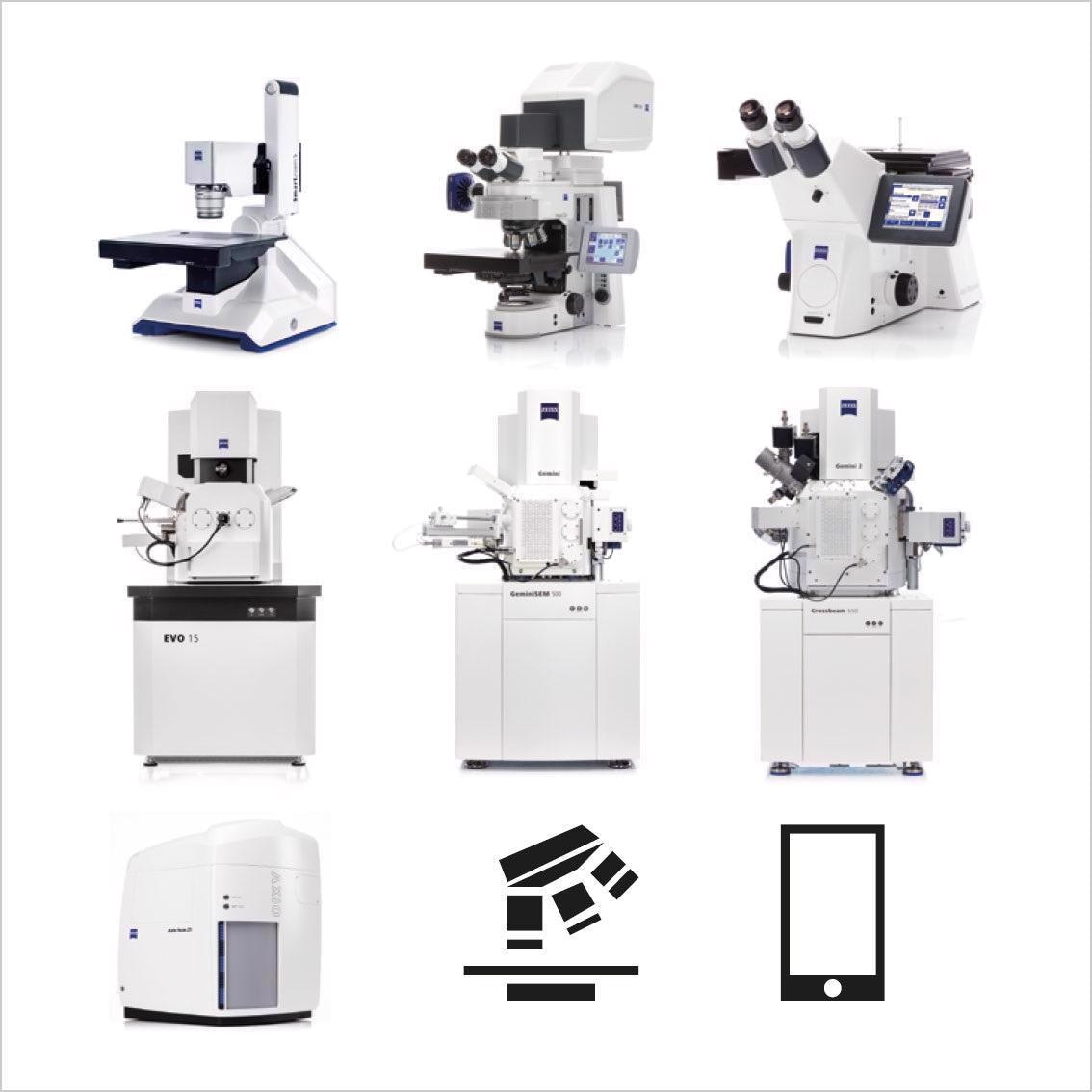
Image Credit: Carl Zeiss Microscopy GmbH
Overlay and Align All Images
- Compatible with all third party imaging technologies
- Simple overview images or complex multi-dimensional images can be loaded from a mobile phone
- ZEISS ZEN Connect stores metadata for longer durations in such a way that external images can follow the well-established Bio-Formats standard
- All image data in context can be aligned and displayed
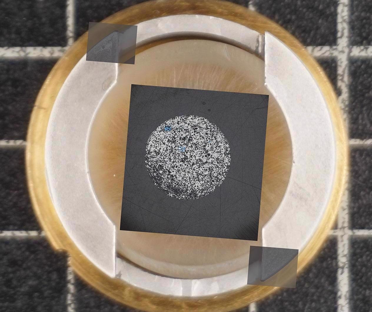
Image Credit: Carl Zeiss Microscopy GmbH
Acquire Overview Images for Easy Navigation
- Mount the specimen on a sample holder from ZEISS or any third party
- Image the sample with any low magnification system or ZEISS stereo microscope
- Use overview images to route and find ROIs
- Identify succeeding images in context while zooming in and out through the borders of imaging technologies and resolution domains
- Adjust the stage to the right position with a click on the overview image and analyze ROIs
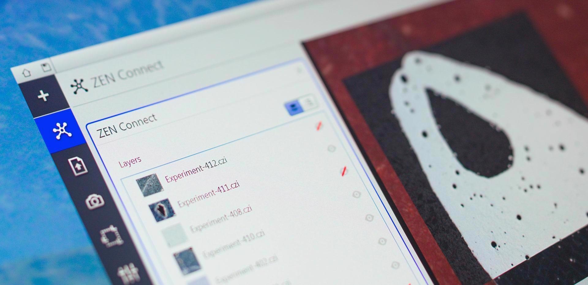
Image Credit: Carl Zeiss Microscopy GmbH
Smart Data Management
- Stay informed about all the steps during the experiments or even months later
- An intuitive label is attached by default to every image once it is saved in the database projects
- Look for imaging parameters and microscope type using the filter function
- Obtain all the images and datasets associated with them
Applications
Material Research in e-Mobility
Permanent magnets are going to decide the future of e-mobility. Start with getting an overview image, then visualize the magnetic domains with a Kerr microscope, and spot reliable magnetic phases. The next steps would reveal further details by increasing resolution from micrometer to nanometer scales.
Based on these results, users can arrive at conclusions and correlate magnetic parameters and sample morphology. It is better to analyze data in a larger context with the best resolution.
Large-scale overview, acquired with ZEISS Axio Imager. Image Credit: T. Schubert, Aalen University, Germany
Magnetic domain structure visualized by Kerr microscopy. Image Credit: T. Schubert, Aalen University, Germany
Correlated high-resolution image acquired with ZEISS Sigma 300 VP. Image Credit: T. Schubert, Aalen University, Germany
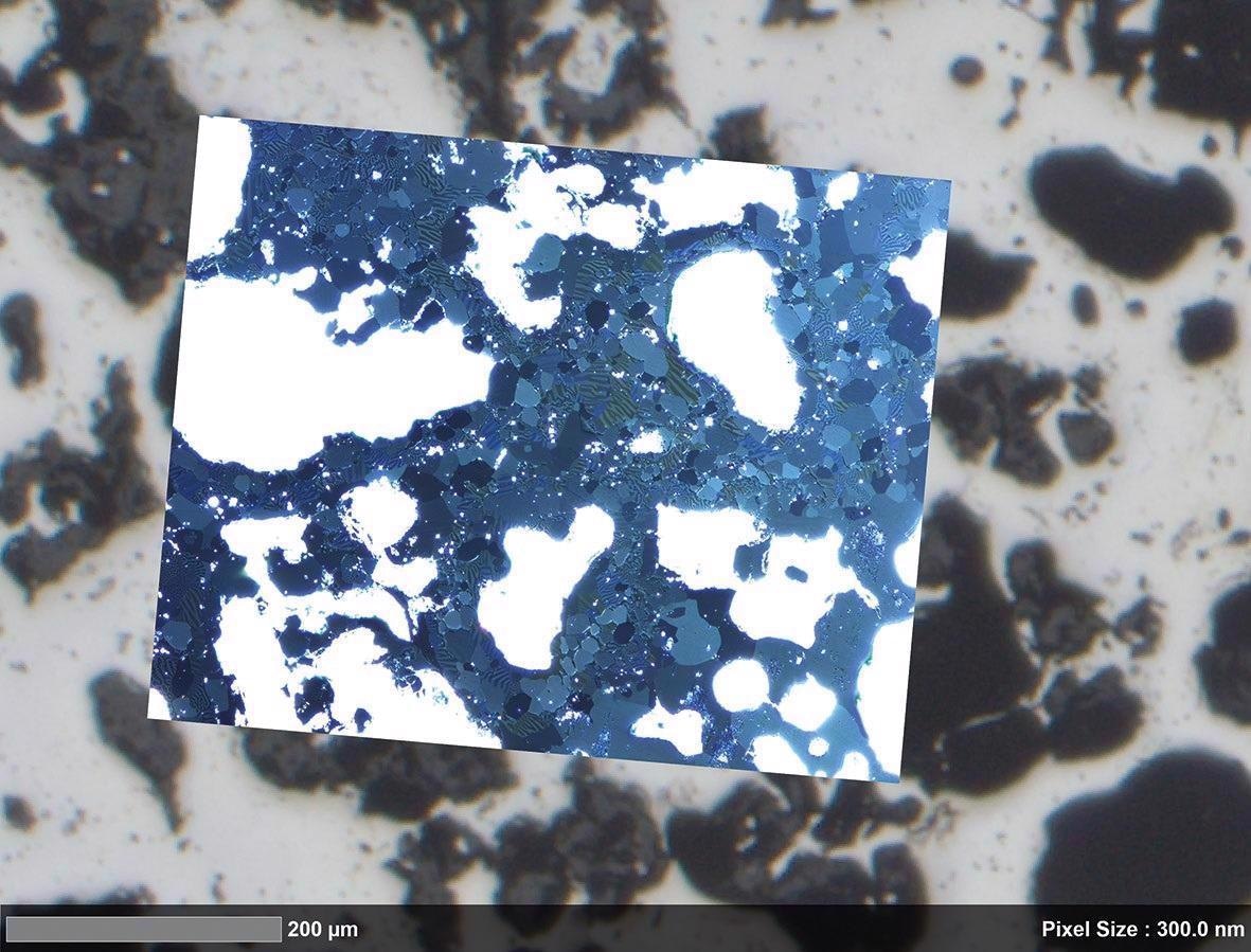
Material Research Oil Recovery, Nuclear Waste Disposal and Carbon Capture and Storage
Examine pore structures of carbonate rocks. Define flow, reaction and transport across various length scales and structural heterogeneity. Use automated light microscopy for a larger picture.
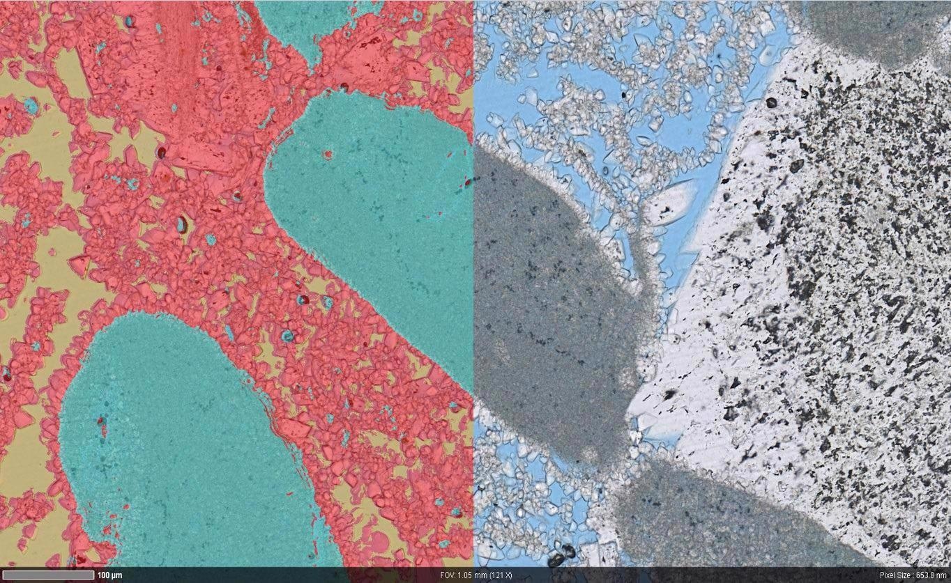
Use ZEISS ZEN Intellesis to classify macro-structure and use different colors, textures and intensities to identify pore structures, microporous grains and solid rock grains. Regions of high-resolution field emission scanning electron microscopy can be identified using the available data. In the context of macroscopic heterogeneity, the difference in nanometer-scale pore structure can be seen.
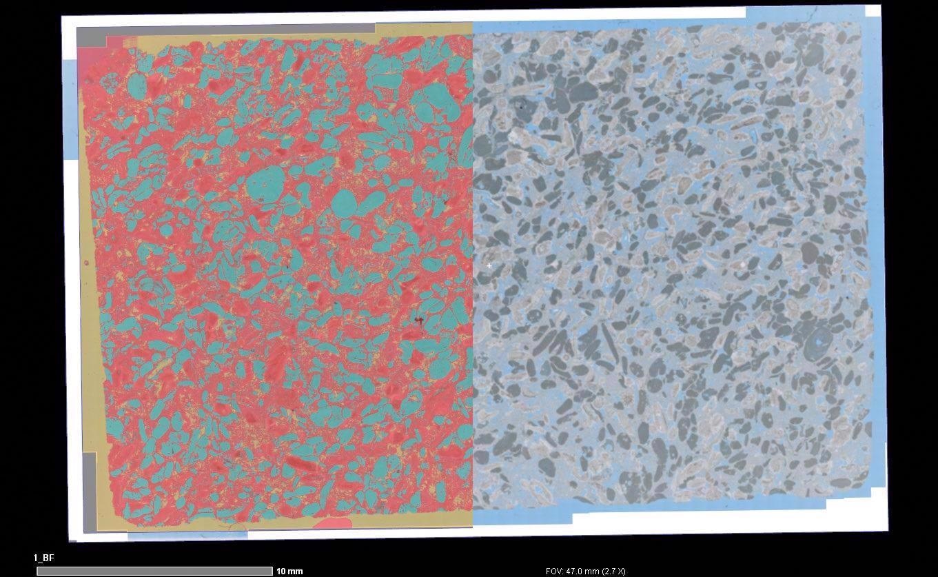
Overlay of macroscopic segmentation (left) and automated light microscopy image (right) showing the microporosity and microporous grains. Image Credit: T. Schubert, Aalen University, Germany
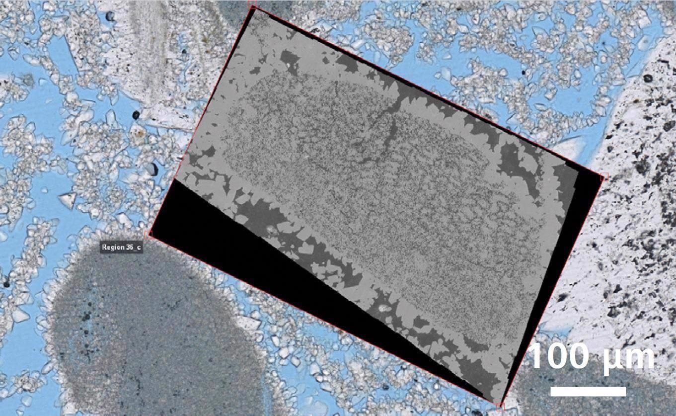
Correlative overlay of high-resolution light microscopy data and nano-scale electron microscopy data. Image Credit: T. Schubert, Aalen University, Germany
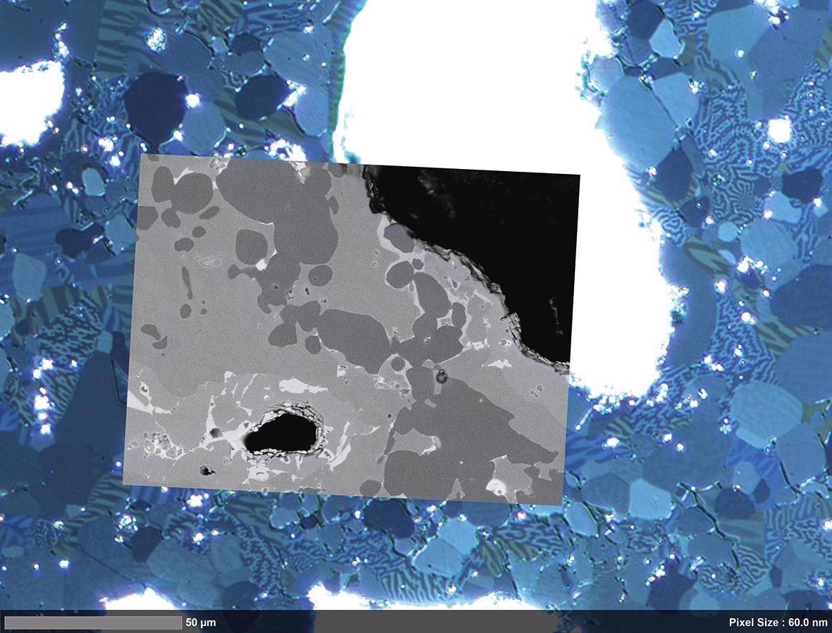
Overlay of segmented (left) high resolution light microscopy data (right) showing the pore structure in more detail. Image Credit: T. Schubert, Aalen University, Germany
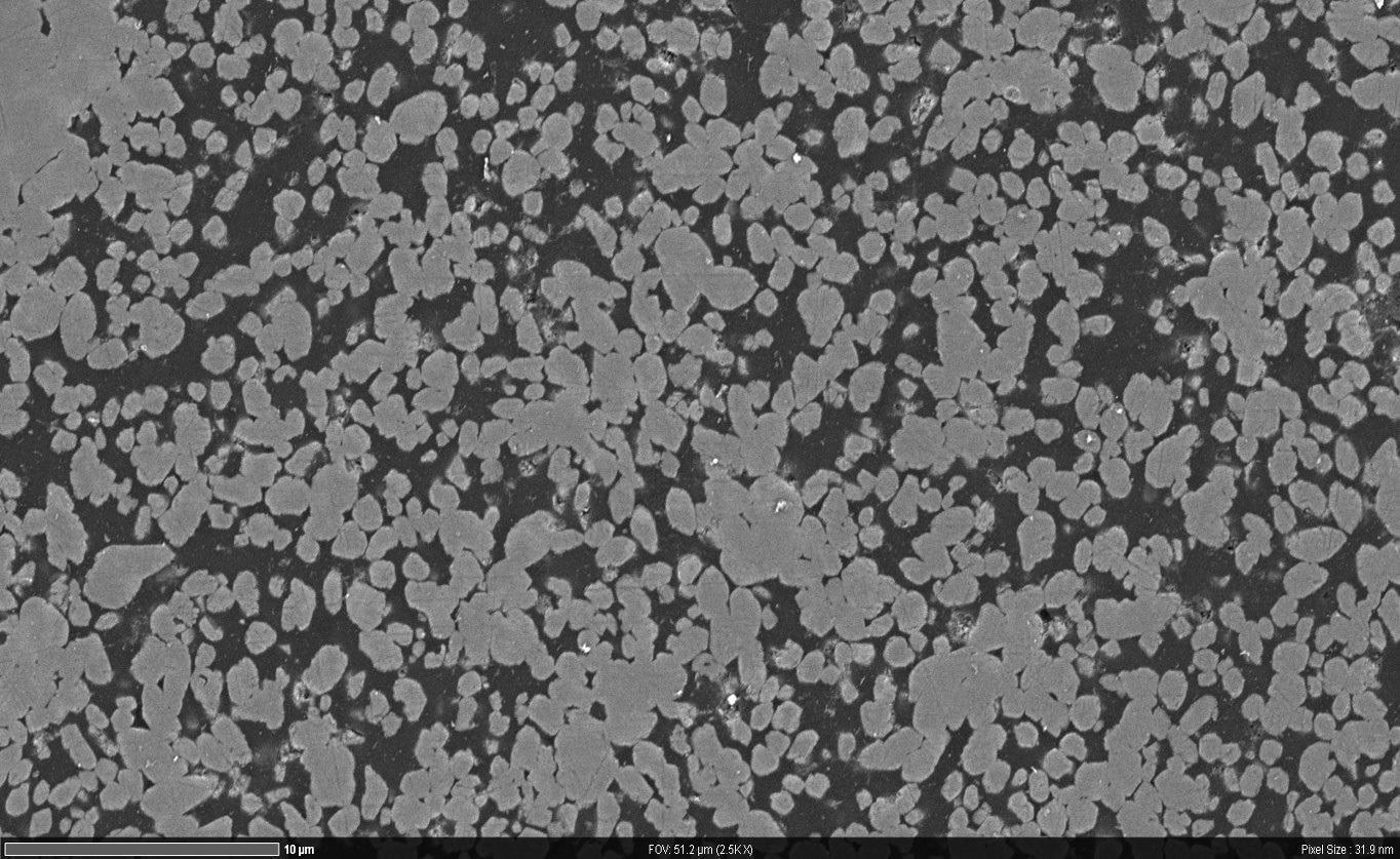
High resolution nano-scale microscope data. Image Credit: T. Schubert, Aalen University, Germany
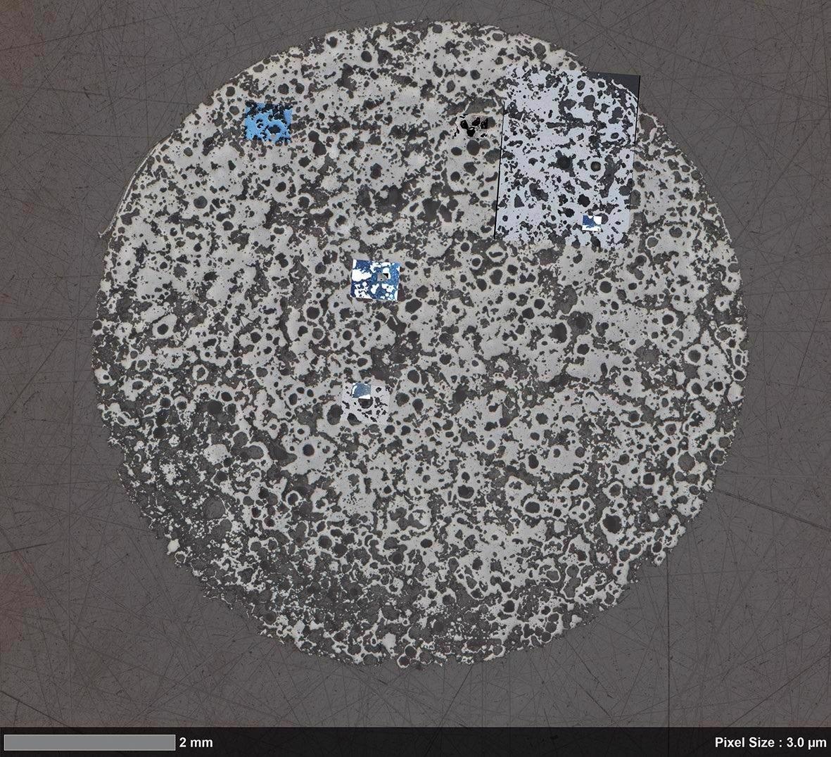
Material Research on Inclusions in Calcium-treated Steel
The formation of detrimental manganese sulfide (MnS) can be controlled by the calcium treatment of steel. This may lead to elongation and hence anisotropy of the steel during rolling.
When modified MnS is used, harder calcium sulfide (CaS) is formed and helps maintain the isotropy of steel. Join both the images to obtain a large-scale view of the entire sample. SEM can be used to obtain a high-resolution image. Change locations to find the specific location of EDS analysis.
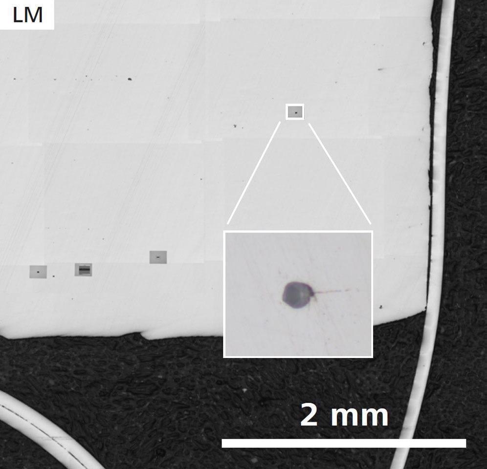
First sample, without Ca treatment: Sample Overview and detail of one inclusion (inset) acquired by ZEISS Axio Observer. Image Credit: T. Schubert, Aalen University, Germany
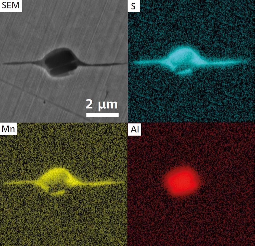
Higher magnification SEM image (ZEISS Crossbeam 550) and EDS maps of an elongated MnS inclusion. Image Credit: T. Schubert, Aalen University, Germany
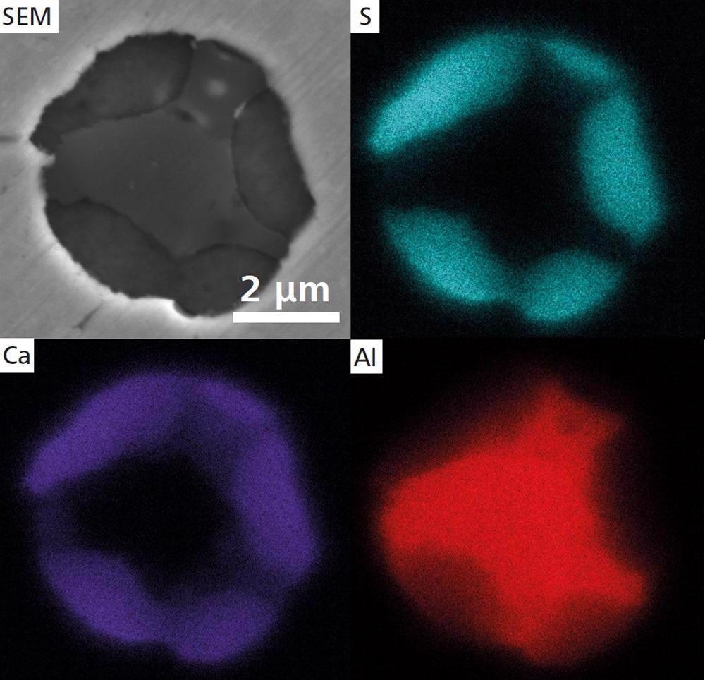
Second sample with Ca treatment SEM image of a globular CaS inclusion and EDS maps. Image Credit: T. Schubert, Aalen University, Germany
Accessories
ZEISS ZEN Data Storage
ZEISS ZEN Data has a central data management system in the laboratory that it is connected to. It enables data acquisition after post-acquisition work and storage of separate images. All the collaborators can benefit from the sharing capabilities and access to data.
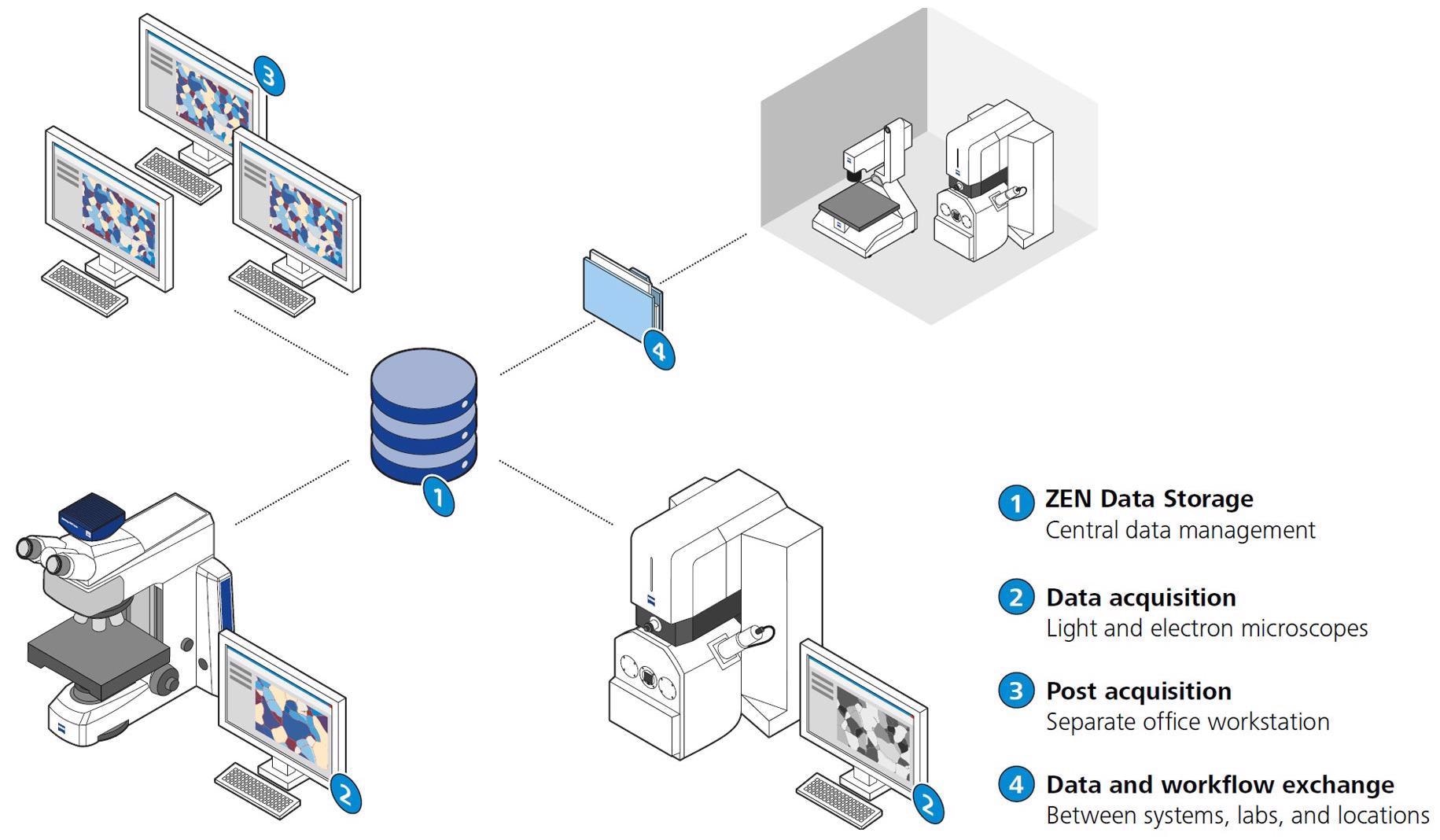
Image Credit: Carl Zeiss Microscopy GmbH
ZEISS Shuttle and Find for Correlative Microscopy
ZEISS Shuttle & Find software module is user-friendly and has a productive workflow to overlay data from scanning electron microscope and light microscope.
To generate a coordinate system within seconds, a specimen holder with fiducial markers can be used. Regions of interest in the sample can be defined with the light microscope. Shift the ROIs in the electron microscope to obtain access to high-resolution imaging and analytics. As a final step, associate the images taken with different microscopical techniques.
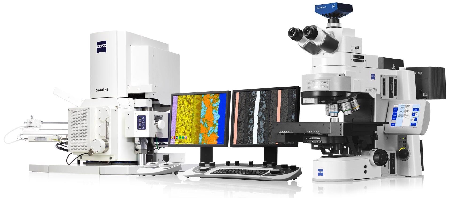
Image Credit: Carl Zeiss Microscopy GmbH
Image Segmentation in Microscopy
The advantages of deep learning are used to easily segment images and get access to real data. Image segmentation is the pivot for all the consecutive image analysis steps.
Deep learning and Python is used by ZEISS ZEN Intellesis to create reproducible segmentation result. Even users who are not experts can train the software, and then the software module will segment images automatically. Hence, it is time-efficient and also reduces user bias.
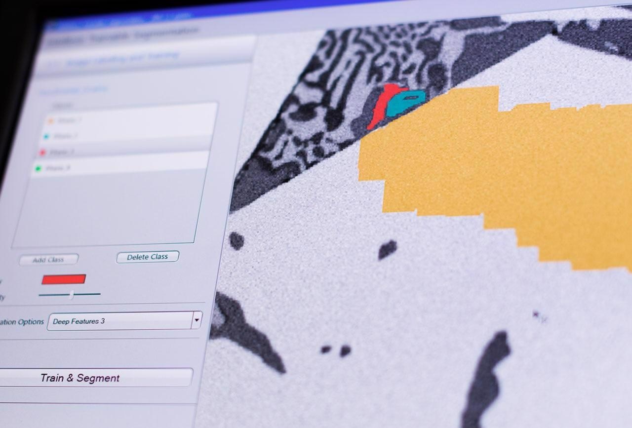
Image Credit: Carl Zeiss Microscopy GmbH