Every aspect of Andor’s new Sona microscope sCMOS camera platform has been engineered from the ground up to optimize performance, drawing the best performance possible from the sensors that have been selected for integration.
There has been a steady increase in interest in back-illuminated sCMOS sensors over the past few years, primarily due to the best-in-class Quantum Efficiency (QE) performance that they deliver. It was a logical step therefore to combine these sensors with best-in-class performance across the rest of the critical areas.
If you have decided to purchase a back-illuminated sCMOS camera, this article makes the case for the Andor Sona models with 7 key reasons why they should be your back-illuminated sCMOS of choice.
The Most Sensitive Back-illuminated sCMOS Available
Sona 4.2B-11 and Sona 2.0B-11 back-illuminated sCMOS models both boast 95% Quantum Efficiency (QE) with market-leading vacuum systems that can cool to -45 °C. The darkcurrent of GPixel back-illuminated sCMOS sensors is comparatively high to the BAE/Fairchild Imaging sCMOS sensors that Zyla and Neo sCMOS cameras use. This heightens the need to deep cool the sensor in order to keep the noise floor as low as possible, i.e. minimizing the camera detection limit.
With its unique vacuum design, Sona uses thermoelectric elements to cool to -25 °C using only the internal fan for heat dissipation. For an even further temperature reduction, Sona can use assisted cooling to drop down to a hugely competitive -45 °C!
Having the most sensitive back-illuminated sCMOS camera carries a vast array of advantages within fluorescence microscopy:
- Reduced laser illumination intensity - keep cells alive throughout study (i.e. suppressing phototoxic effects) and also limit dye photobleaching
- Reduced fluorophore concentrations - maintaining accurate physiology in living specimens
- Lower exposure times - follow faster processes
- Better SNR with TIRF and confocal low light modalities - better image clarity with techniques that reject out of focus photons.
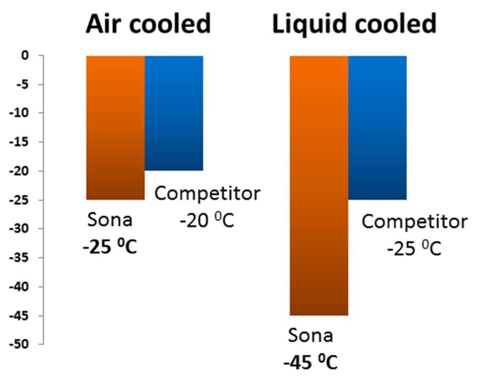
Figure 1. Air (fan) cooled and liquid cooled performance of Sona back-illuminated sCMOS models, versus the nearest competitive backilluminated sCMOS camera type.
Largest Field of View Available: Sona 4.2B-11
Compared to other, competing back-illuminated sensors that also use the same GPixel GS400 BSI sensor type, the Sona 4.2B-11 model offers the largest field of view solution. The Sona 4.2B-11 is native F-mount and is compared below against a similar model, or “Competitor A”, a camera using the same sensor but cropped down to 1608 x 1608 pixel format.
This camera avoids sensor glow issues at the sensor edges by cropping the sensor down. However, Sona 4.2B-11 uses a unique Anti-Glow Technology approach that allows the entire native 2048 x 2048 of the array to be harnessed. The 62 % increase field of view advantage offered by Sona 4.2B-11 is seen in Figure 2.
Having as high a field of view as possible with Back-illuminated sCMOS cameras is crucial for a range of studies, including:
- Developmental biology – capture whole embryos, e.g. Zebrafish
- High Content Screening – capture larger fields of cells, increase information content
- Tissue Cultures – minimize stitching, maximize throughput
- Organoids – unravelling cell connectivities
- Gene Editing – screening large cell cultures for cells whose genomes have been successfully edited
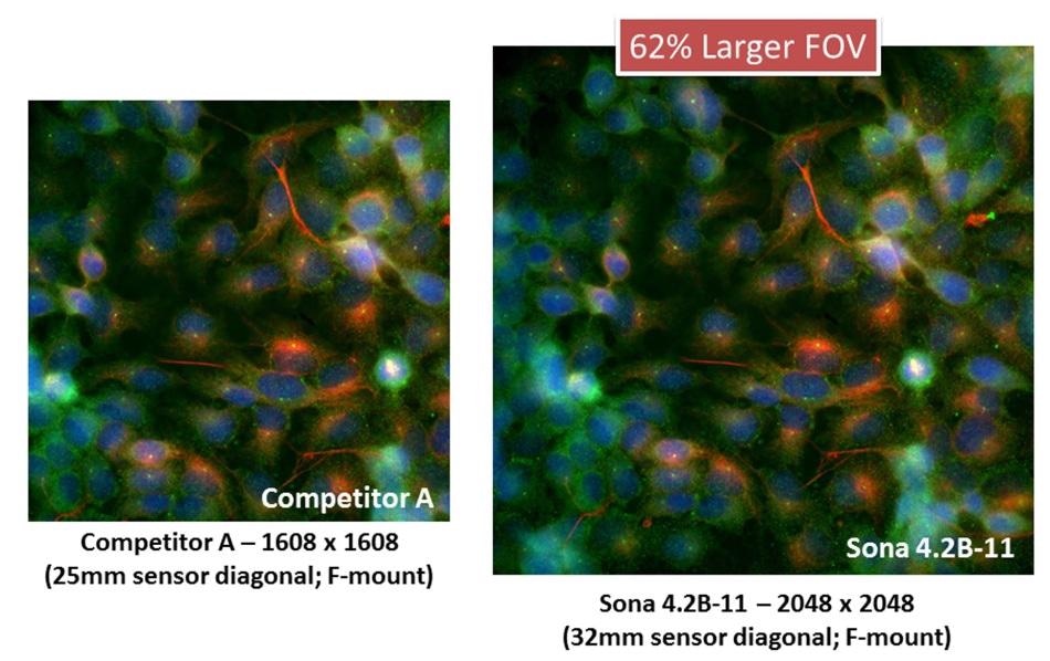
Figure 2. “F-mount competitive solutions” – Field of View comparison between Sona 4.2B-11 and a competitor Fmount camera, utilizing the same GS400B back-illuminated sCMOS sensor but restricted to 1608 x 1608 max resolution. Captured using a Nikon Ti2 with 60x objective and integrated 1.5x tube lens. The Sona 4.2B-11 has 62% more active pixels and offers a compelling field of view solution.
Enhanced Mounting Flexibility: One camera, Multiple ports
Sona 2.0B-11
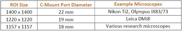
The Sona 2.0B-11 is native C-mount and can be interfaced with various microscope C-mount port diameters, up to 22 mm. The 1400 x 1400 full array size of this model is suited to modern 22 mm C-mount ports and makes full use of the available field of view from this common mount type. If smaller microscope port sizes are they only ones available, there are other pre-configured, centrally positioned ROIs are available.
Table 1. Pre-configured ROIs of the C-Mount Sona 2.0B-11 model, shown alongside the corresponding microscope Port Diameter / Field Number for which they are optimized.
Another option for the use of smaller ports is the Andor Magnifying Coupler Unit. This is a complimentary coupler that can readily connect to the port, utilizing the full 1400 x 1400 array size and expanding the image available from the microscope onto the larger sensor area. A 2x coupler also makes it possible for Nyquist resolution utilizing a 60x objective, which then further optimizes the on-sample field of view.
Sona 4.2B-11
The superior flexibility of the Sona 4.2B-11 allows use with all ports. By coupling the full array of 32 mm with the Nikon Ti2 F-mount and in-built 1.5x tube lens, unparalleled on-sample field of view and superb uniformity is obtained
It is also possible to use the Andor Magnifying Coupler Unit with this system, for access to a large variety of modern research fluorescence microscopes and corresponding ports, gaining an additional 2x magnification onto the large sensor area of the Sona 4.2B-11. Since the image is magnified 2x onto a 32 mm diameter sensor area, the Magnifying Coupler Unit can be used with any port that offers an image output of 16 mm or greater; this applies to the vast majority of available ports.
Finally, while the Sona 4.2B-11 is pre-installed as an F-mount system, the camera can be easily converted to either C-mount or T-mount, using accessories that can be ordered at the same time as the platform.
Something to note is the Andor Magnifying Coupler Unit is T-mount at the camera attachment end, making it necessary to convert the camera to a T-mount before they can be used together. This brings a benefit in that the interface between the two will be completely light tight- light leak being sometimes otherwise associated with F-mount side port usage.
Figure 3. The Andor Magnifying Coupler Unit (orderable with Sona models)
Superior Quality & Longevity – UltraVac
Vacuum technology affords two main advantages to the system. While primarily working to minimize the noise floor, Andor’s vacuum sensor enclosure also provides longevity benefits that should not be overlooked.
Reason 1: Sensor Protection
Without protection, back-illuminated silicon sensors are susceptible to attack from moisture, hydrocarbons and other gas contaminants that cause a steady decline in performance, including QE decline. UltravacTM is Andor’s proprietary vacuum enclosure technology, with minimized out-gassing, offering the best level of protection for the sensors.
Reason 2: No Re-Backfilling of Sensor Enclosure Required
Without a vacuum enclosure, cameras instead have to use a method called back-filling, whereby the sensor enclosure is flushed with a dry gas to remove contaminants which then remains as a positive pressure of dry gas, separated from the external atmosphere by only O-ring seals.
As these seals are not completely impenetrable by atmospheric water and gas, over time they will enter the sensor enclosure and compromise the system. This results in loss of cooling capability and often moisture condensing on the sensor, making it necessary for the entire camera to be sent back to the factory for repair, re-backfilling and resealing, often outside of the warranty period. By using a hermetic vacuum seal, the UltraVacTM system prevents any and all gas ingress from the outside environment, with the vast majority of cameras never losing any cooling performance.
Sona is the only back-illuminated sCMOS camera commercially available that has the advantage of a vacuum sensor enclosure. Andor have over 25 years of expertise in vacuum sensor technology, and they hold it as one of their core technology strengths, having a fantastic track record of vacuum integrity and associated camera longevity.
Higher Quantitative Accuracy
Sona 4.2B and Sona 2.0B are both supported with Extended Dynamic Range functionality, across a 16-bit data range. Harnessing innovative ‘dual amplifier’ sensor architecture, the maximum pixel well depth and the lowest noise can be accessed simultaneously, allowing quantification of extremely weak and relatively bright signal regions in one snap. This feature is excellent for accurately visualizing and quantifying those samples that have both weak and bright regions, neurons for example.
Andor have implemented enhanced on-head intelligence to achieve best in class quantification accuracy, delivering linearity of >99.7%.
There are many applications that require accurate quantitative information over only structural detail; any measurement where intensity correlates to quantity or concentration will benefit from superior linearity. This can refer to physiological parameters such as calcium, pH, cAMP etc. with single and dual wavelength dyes.
Gene expression analysis with fusion proteins demands superb quantitative accuracy, where intensity is directly correlated to concentration. FRET analysis also uses the relation of intensity to concentration (by chelation e.g. calmodulin) as well as distance or co-localization at the nanometer scale. Localization Super-Resolution Microscopy can also be negatively affected by poor linearity because the Gaussian fit could become skewed.
Fast Speed Mode
Both Sona 4.2B and Sona 2.0B offer fast frame rate capability, making them perfect for following dynamic cell processes such as ion signaling, cell motility and blood flow, and eliminating image smear. If a further boost to frame rate is needed, Region of Interest (ROI) and 12-bit readout mode can both be utilized.
For those applications where speed is needed over range, Sona 4.2B and Sona 2.0B are each architected to offer both 16-bit and 12-bit modes. At the cost of wide dynamic range, selecting 12-bit will accelerate frame rate by 2x, useful for imaging fast processes using low light modalities such as spinning disk confocal or TIRF.
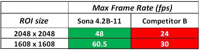
Table 2. Not all commercial cameras utilizing the GS400 sensor offer this boosted speed capability. Maximum frame rates of Sona 4.2B-11 are shown here versus a competitive camera using the GS400 sensor that does not offer this 12-bit fast speed mode.
Maximizing speed with back-illuminated sCMOS cameras expands capabilities in a range of studies, including:
- Ion Signaling - Follow fast calcium wave propagation and calcium sparks with maximum temporal dynamics, with further accelerations possible through use of ROIs. For elongated smooth muscle cells, rectangular ROIs can be used (up to the full width of the sensor) without imposing a speed compromise.
- Cell Motility – Speed capability is critical for following cell movement, e.g. sperm cell dynamics.
- Intracellular transport – Fast frame rates can be important to follow intracellular transport dynamic, including membrane dynamics.
- Blood flow – increased speed is critical for perhaps one of the most temporarally challenging applications.
- Localization super-resolution – Back-illuminated sCMOS is increasingly popular for localization super-resolution, as the higher QE yields higher SNR and therefore better localization accuracies. However, many raw images have to be rapidly acquired for a single super-resolved output image: boosted speed is critical, especially if live cell super-resolution is the true goal.
Enhanced Flexibility
Designed with the requirements and challenges that the modern research environment brings, the Sona platform has a camera that is inherently flexible and can be adapted across an array of setups and experimental configurations. The following areas of flexibility are built-into the Sona platform:
- Air and liquid Cooling –the liquid cooling operation of the Sona not only maximizes sensitivity in extreme low light conditions, using it over fan assisted cooling can also be beneficial in experiments that are particularly vibration sensitive, such as electrophysiology experiments or combined optical/AFM set-ups.
- 16 and 12-bit modes – The 16-bit mode is ideal for imaging samples that have both weak and bright signal regions, with its High Dynamic Range mode. The 12-bit Fast Speed mode can be utilized to increase available frame rate 2x using any selected ROI size, superb for adapting to low light experiments that require excellent temporal resolution such as ion signaling or blood flow imaging.
- Adapt to multiple ports or objectives - As covered in more detail earlier, both Sona 4.2B-11 and Sona 2.0-11 models can be readily adapted for use over a wide range of microscopes with varying port sizes and coupling attachments (e.g. F, C and T-mount). This allows the camera to be easily swapped through multiple experimental set-ups. Also, the on-sample field of view can be maximized using the Andor Magnifying Coupler Unit to adapt the 11 µm pixel sensor to lower magnification objectives.
- Flexible pixel binning – The Sona models have on-camera flexible pixel binning functionality, user definable to 1 pixel granularity. Heightened binning flexibility can be useful when resolution can be sacrificed in favor of enhanced photon collection area per pixel - e.g. extremely lower light bioluminescence experiments.
- Timestamp – The Sona platform can timestamp an image with an accuracy of 25 nanoseconds. Accurate timestamps are vital where precise knowledge of frame time impacts temporal dynamic analysis, and are especially important for fast events, where computer and interface latencies need to be considered. Areas include signaling cascades, vesicle trafficking, lipid dynamics, synaptic re-modelling, action potential studies using opto-genetics and opto-physiology. Timestamps can also be useful for FRAP Analysis, facilitating the estimation of diffusion rates.

This information has been sourced, reviewed and adapted from materials provided by Andor Technology Ltd.
For more information on this source, please visit Andor Technology Ltd.