At the National Research and Development Institute for Textiles and Leather (INCDTP-ICPI) in Bucharest, researchers at the Advanced Research for Cultural Heritage Laboratory (ARCH Lab) employed the LTS120 Peltier system by Linkam to analyze the degradation of collagen fibers in historical leather and parchment1.
At the University of the Basque Country, researchers at the Department of Analytical Chemistry utilized a Linkam THMS600 to apply Raman spectroscopy for the investigation of the phase transition of historical paintings based on plattnerite [ß-PbO2 lead (IV) oxide]2.
These case studies are two of many that demonstrate the performance of temperature-controlled microscopy in a broad range of scientific disciplines, from metallurgy and materials to oil and gas, life science, food research, and space exploration.
Whether it is a canvas oil painting from the 15th century, tools or clothing from prehistoric times, a handwritten document on parchment from ancient Rome, or even a mummy, many marvel at how well these artifacts have been preserved for hundreds and even thousands of years.
Historians and scientists have paid significant attention to understanding the degradation of archaeological and historical artifacts and what steps can be taken to preserve them more effectively.
A team of researchers from the ARCH Lab, INCDTP-ICPI, Bucharest, collaborated with the National Museum of Romanian History and the Institute of Natural Sciences and Technology in the Arts, Academy of Fine Arts, Vienna, to evaluate and monitor the physical-chemical changes of parchment as a result of photo-oxidative aging.
Non-invasive Fourier transform infrared spectroscopy in attenuated total reflectance acquisition mode (FTIR-ATR) was applied, which is suitable for natural and biological samples, along with colorimetric measurements and Raman spectroscopy, and the Micro-Hot-Table (MHT) technique.
MHT is a micro-invasive technique that is commonly employed to determine the shrinkage temperature (Ts) of collagen-based archaeological and historical materials (predominantly parchment and leather). The value of Ts relies on the fibrous collagen’s structural stability, and, therefore, on its degree of deterioration.
Investigations were carried out utilizing MHT equipment that included a Linkam LTS120 temperature control Peltier stage with an added stereomicroscope and an automatic heating rate adjustment system.
A digital camera fixed onto the microscope was used to record the shrinking motion (as shown in Figure 1). The basic technique in combination with equipment that provides advanced analysis facilitates in situ investigation and the characterization of collagen-based artifacts.
The experiment in this work was carried out to study the damaging effects of mixed light-thermal aging on collagen in the solid phase, as found in parchment. If we better understand how collagen deteriorates in a particular artifact, we can get helpful insight into its manufacture, how damaged it is (see Figure 1), as well as how best to formulate conservation treatments and apply appropriate storage and handling conditions to preserve it.
We have been extensively using the MHT method since 2006 and are now strongly advocating a more analytical use of this method when analyzing both new and artificially aged samples, and ancient materials, by including the analysis of all shrinkage parameters to limit the risk of over or underestimating the conservation (damage) condition of parchment/leather.
Project Coordinator, Arch Lab and Project Director, INCDTP-ICPI
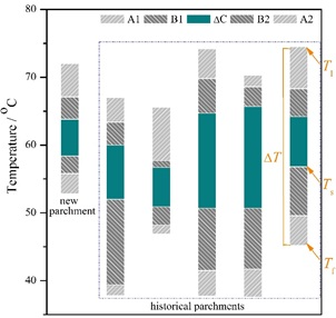
Figure 1. Graphical presentation of the shrinkage intervals and temperatures for some historical documents on parchment belonging to the Collection “Royal Chancellery of the Stephen the Great (1457-1504)” from the Romanian Academy Library highlighting the ability of MHT method to differentiate the parchments depending on their degree of damage. Figure reproduced from reference 1 in accordance with Creative Commons Attribution License.
“The FTIR-ATR and Raman techniques coupled with MHT method provided qualitative results that allowed us to characterize the aging pattern of collagen in parchment exposed to light irradiation at 52 °C and 30% relative humidity over time.”
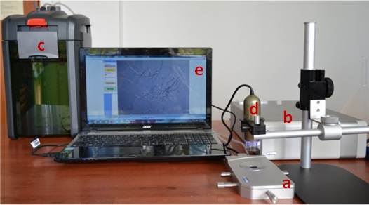
Figure 2. MHT equipment for in situ analysis of shrinkage activity used at ARCH Lab, INCDTP-ICPI, Bucharest. (a) micro heating plate (Linkam LTS 120); (b) temperature controller; (c) water circulator; (d) digital microscope; (e) computer with image MHT software.
The LTS120 Peltier stage is a temperature control stage that is simple to use and designed for use with optical microscopes.
A 40 mm x 40 mm Peltier element is used in the instrument to control the sample’s temperature ranging from -25 to 120 °C with rates of up to 30 °C per minute and a 0.1 °C accuracy. It includes features for humidity control, electronic connections, and gas purging. The sample can be moved 15 mm in both X and Y directions and can be fixed onto a regular microscope slide.
Plattnerite is a by-product produced through the deterioration of lead-based pigments, which have been commonly used to create artwork for centuries. The creation of plattnerite and additional lead compounds is thought to be the cause of an unwanted blackening in historical artworks.
The restoration and analysis or laser cleaning of historical artwork can be assisted by an improved understanding of plattnerite formation. At the University of the Basque Country, the research team used a Raman Spectrometer coupled with a THMS600 temperature-controlled stage to determine the temperature range at which the phase transitions of lead dioxide occurred by heating commercial plattnerite samples and increasing the temperature of the cells.
Raman analysis was used to demonstrate the gradual deterioration of plattnerite and the production of secondary products. The team discovered that pigments from aged painting samples behaved in a different manner to those found in commercial samples under the laser’s influence.
The THMS600 freezing–heating stage can be operated over a temperature range of -196 to +600°C and it enabled us to verify that the first phase transformation of plattnerite takes place at a temperature between 365 and 370°C. The stage allowed us to obtain a gradual transformation of lead-based compounds from red lead into litharge and massicot (types of lead oxide minerals), generated by the thermal plattnerite degradation (see Figure 3).
However, we also found that the degradation process was reversible, since only the use of the laser beam, even if used at maximum power, was not enough to obtain a stable form of massicot. The intermediate products of the decomposition of plattnerite were evident when the sample was analyzed again at low laser power, after cooling.
The temperature-controlled stage allowed us to see the Raman spectra belonging to the different phase transitions, which had not been possible via varying the laser power alone. Again, when the thermal input disappears, the return to the a-PbO (litharge) compound is observed. Our findings indicate that, in the presence of lead compounds, it is important to use a low laser power during Raman measurements, on mural paintings’ surfaces.
Dr Juan Manuel Madariaga, A member of the Research Team
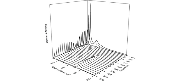
Figure 3. 3D representation of Raman spectra shows the phase transition from red lead to litharge and then massicot, with increasing temperature (from 500°C to 600°C) using the THMS600. Figure reproduced from reference 2 in accordance with Creative Commons Attribution License.
A number of once-unknown key clues in historical research are now being identified through the investigation of artifacts, manuscripts, and documents, which have been rarely opened, exhibited or read.
For example, the spread of production techniques, the interactions of the materials within the environments that they were used, and the origin of the materials are all elements of knowledge that must be grasped to understand the complete picture of a historical artifact.
About Linkam Scientific Instruments
Linkam manufactures and develops a wide array of temperature-controlled stages from high to cryo temperatures for both end-users and OEM, and can offer tailored solutions.
The stages are utilized in combination with light microscopes and a broad selection of analytical methods, including FTIR, Raman, WAX/SAX and additional X-ray techniques to characterize and visualize the properties of materials.
Thousands of laboratories across the globe utilize stages by Linkam. The THMS600 is one of the most commonly used freezing and heating microscope stages on offer, and is employed for a range of applications where high freezing/heating rates and 0.01°C stability and accuracy are necessary.
Samples are efficiently characterized by heating them to within a few degrees of the desired temperature with minimal overshoot and at a rate of up to 150°C/min. The heating is then slowed down to a few tenths of a degree per minute to analyze changes to the sample in detail. Linkam is an industry leader in temperature-controlled microscopy.
References and Further Reading
- Studies on the effects of mixed light-thermal aging on parchment by vibrational spectroscopy and micro hot table method, Cappa et al. Herit Sci (2020); Micro differential scanning calorimetry and micro hot table method for quantifying deterioration of historical leather. Carsote and Badea, Herit Sci (2019).
- Use of temperature-controlled stage confocal Raman microscopy to study phase transition of lead dioxide (Plattnerite), Costantini et al. Minerals (2020).
This information has been sourced, reviewed and adapted from materials provided by Linkam Scientific Instruments.
For more information on this source, please visit Linkam Scientific Instruments.