The Helmholtz Center for Materials and Energy in Berlin, Germany (HZB, originally known as Hahn-Meitner Institute, HMI) has developed a novel technique for the cross-sectional sample preparation for transmission electron microscopy (TEM).
This new preparation method – primarily applied to thin-film stacks of solar-cell devices – involves the mechanical and argon (Ar) ion polishing of cross-section samples that have been fabricated via the face-to-face fixing of two thin-film stacks with epoxy glue.
The T-Tool is a useful implement employed in the mechanical polishing of cross-section samples. This tool – introduced by H. Zang in 19981 – is similar to the more widely known Tripod.2
The T-Tool was further developed and refined by Peter Schubert-Bischoff at HMI/HZB and has since been utilized by a wide range of labs across the globe. Figure 1a displays the T-Tool (HMI style) with an attached glass cylinder.
The mechanical polishing procedure sees the cross-section sample being lapped on diamond foils using different sizes of diamond particles. A stabilizing metal ring is then glued on top of the polished sample, and carbon fibers are placed across the cross-section’s interface region (Figure 1b).

Figure 1. a) Photography of the T-Tool developed at HMI/HZB, with a glass cylinder attached to it, on top of which the specimen is clued by use of hot wax. b) Light micrograph of the inner part of a Mo support ring, showing the interface region (black vertical line in the middle of the image) with the glass substrates of the thin-film stacks on either side, as well as carbon fibers glued across the interface region of the cross-section. Image Credit: Gatan Inc.
These carbon fibers act as screens for the ion beams during the Ar ion milling step of the preparation procedure, allowing the regions in between the carbon fibers to be preferentially milled.
Using this approach results in the generation of several positions where the sample is transparent to the electron beam (Figure 2a, red ellipses).
The sample can be Ar ion polished multiple times, maximizing the number of positions at which TEM analyses may be conducted for each sample.
As a TEM study typically seeks to analyze a specific position in a sample, the PIPS II system’s X, Y stage allows the Ar beams to be aligned to a region of interest. This allows individual regions between the carbon fibers to be milled effectively (Figure 2b).
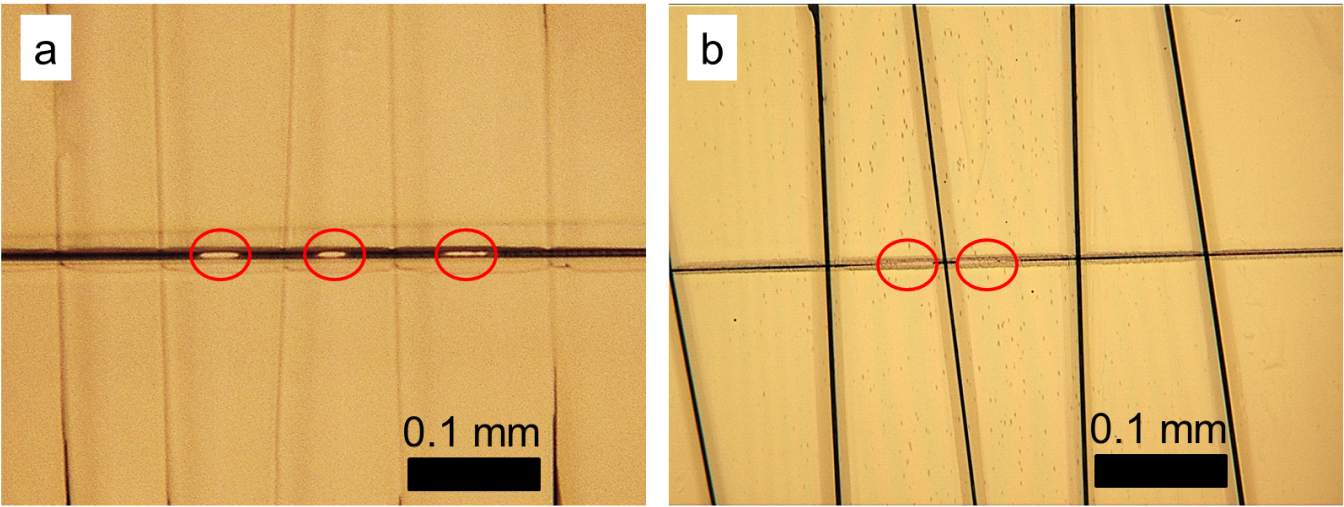
Figure 2. a) Photography of a cross-section specimen after ion milling using a broad Ar ion beam, resulting in several holes in between the carbon fibers. b) The PIPS II system permits alignment of the Ar beams in such a way that holes form only within a small region of interest. Image Credit: Gatan Inc.
This improved approach to sample preparation was employed in the preparation of cross-section samples of a ZnO/CdS/Cu(In,Ga)Se2/Mo glass thin-film solar cell.
When working with this material system, a high standard of sample preparation is necessary to accommodate the mechanically unstable interface between Mo and Cu(In,Ga)Se2.
As the ion milling of Cu(In,Ga)Se2 is frequently incongruent, Cu agglomerates (Figure 3a, red circles) may form on the sample’s surface.3 The application of liquid N2 during Ar ion milling in the PIPS II system (Figure 3b) can significantly reduce the occurrence of these agglomerates. This resulted in a working temperature of -40 °C.

Figure 3. a) Bright-field TEM image of a ZnO/CdS/Cu(In,Ga)Se2/Mo solar-cell stack. Cu agglomerates on the Cu(In,Ga)Se2 surface are highlighted by red circles. b) No Cu agglomerates visible on a specimen prepared using optimized Ar ion beam parameters and liquid N2 cooling in the PIPS II system. Image Credit: Gatan Inc.
The use of optimized parameters and sample preparation techniques has been shown to enable the structural properties of ZnO/CdS/Cu(In,Ga)Se2/Mo solar-cell stack’s individual layers - and their interfaces - to be analyzed at high resolution (Figure 4).
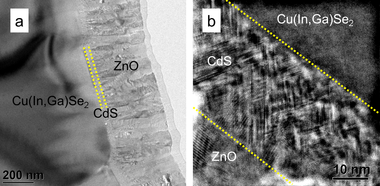
Figure 4. Bright-field (a) and high-resolution TEM image (b) of the ZnO/CdS/Cu(In,Ga)Se2 stack prepared using liquid N2 cooling in the PIPS II system during Ar ion milling. Structural properties such as extended defects and orientation relationships between the CdS and Cu(In,Ga)Se2 layers can be analyzed at the sub-nanometer scale. Image Credit: Gatan Inc.
References
- H. Zang, Thin Solid Films 320 (1998) 77-85.
- J. B. Benedict, R. Anderson, S. J, Klepeis, M. Chaker, Specimen Preparation for TEM of Materials II, Mat. Res. Soc. Symp. Proc. 199 (1990) 189-204; J. B. Benedict, R. Anderson, S. J. Klepeis, Specimen Preparation for TEM of Materials III, Mat. Res. Soc. Symp. Proc. 254 (1991) 121-140.
- D. Abou-Ras, B. Marsen, T. Rissom, F. Frost, H. Schulz, F. Bauer, V. Efimova, V. Hoffmann, A. Eicke, Micron 43 (2012) 470-474
Acknowledgments
Produced from materials originally authored by Daniel Abou-Ras, Ulrike Bloeck, and Peter Schubert- Bischoff from the Helmholtz Center for Materials and Energy, Berlin.

This information has been sourced, reviewed and adapted from materials provided by Gatan Inc.
For more information on this source, please visit Gatan Inc.