Microplastics are a growing problem in the world’s oceans, but how do you obtain quick and useful measurements in-situ, and at the depths needed to completely understand distribution, flow, and ocean life consumption? Filtration-based methods that use Fourier-transform infrared spectroscopy (FTIR) and Raman microscopy are well-proven, however, they work best when taking surface measurements, where microplastic densities are higher.
A collaborative team of researchers from Japan and the UK is developing a hybrid, compact system using holography and Raman spectroscopy. The solution monitors particle size, shape, and material in-situ – offering promise for monitoring those depths where sea life exceeds the number of plastics.
The Plastics Problem Runs Deep
Plastic pollution extends beyond the surface and is not confined to discrete items such as plastic bags or large accumulations like the Pacific Garbage Patch. As each of these larger sources of plastic pollution deteriorates, they generate secondary pollution called microplastics, particles less than 5 mm in size that sink and reach deep ocean depths.
Microplastics are ingested by marine life, infiltrating the food chain, which many species depend upon. In-situ monitoring data is required to properly evaluate the extent, distribution, and changes in microplastic buildup in the ocean over periods of time, specifically as a function of depth.
This data supports studies on how organisms and microplastics interact, and can be used to enhance vertical transport models. In the future, it will serve as a guide for remediation efforts and enable effectiveness assessments.

Image Credit: Wasatch Photonics, Inc.
Seeing Microplastics in Seawater
A number of the existing “collect & separate” techniques for identifying microplastics are focused on sampling surface waters and necessitate filtration to sufficiently concentrate the sample for detection. Due to the weak signal, the spectra acquired via Raman spectroscopy are data-rich and much easier to acquire with a bulk sample.
The density of microplastics in deeper waters is much lower than at the surface, and the presence of native microscopic marine life of similar dimensions can be rich. This presents two challenges for Raman-based detection:
- Ensuring the signal required is achieved
- Scanning the appropriate amount of seawater in the presence of undesirable background or ‘noise’.
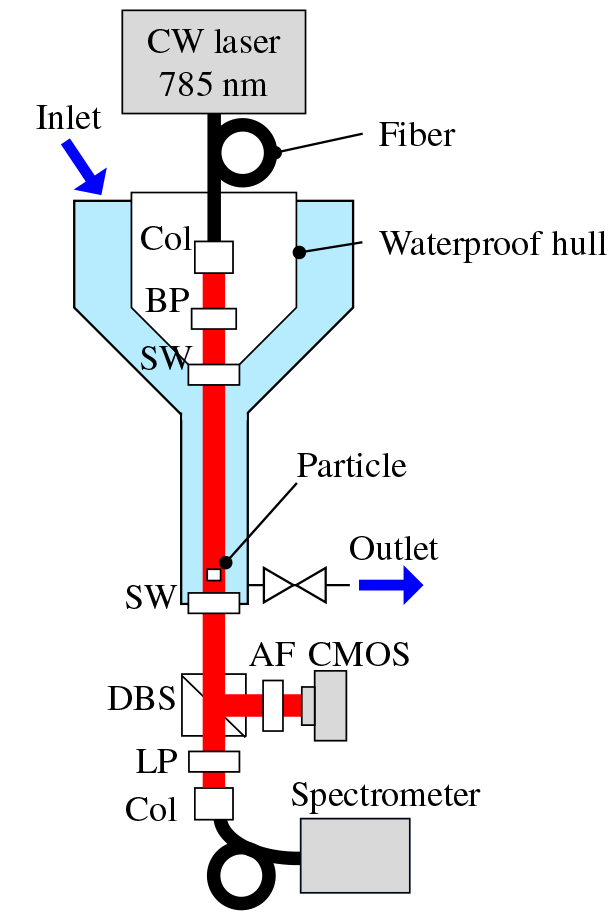
Figure 1. Hybrid monitoring system, experimental setup: Col, collimator; BP, bandpass filter; SW, sapphire window; DBS, dichroic beamsplitter; AF, attenuation filter; and LP, longpass filter. Reprinted with permission from [1] © The Optical Society
To help overcome these challenges, the collaborative team of researchers led by the Japan Agency for Marine-Earth Science and Technology (JAMSTEC), the University of Tokyo, the University of Southampton, and the University of Aberdeen have developed a compact but sensitive hybrid flow-through detection system with the capacity to characterize individual particles.
The system depends on digital holography to pre-screen particles of interest by size and shape to establish if they are man-made or natural. Raman spectroscopy can then be carried out on a selective basis, increasing the system’s throughput. This would enable continuous and efficient in-situ monitoring even at depths where microplastic particles are less common.
The team developed a dual-purpose system with a single 785 nm laser as an illumination source with imaging and Raman spectroscopy capabilities to prove the concept.
After passing over the sample flow channel, part of the transmitted laser light is redirected by a beam splitter towards a fast CMOS camera for digital holographic imaging; the rest is left up to Raman spectroscopy.
The team was keen to assess whether Transmission Raman, routinely used for pharmaceutical analysis, could help distinguish microplastic particles in-situ without physically trapping them on a membrane.
A compact Wasatch Photonics WP 785 ER spectrometer is employed for the material analysis, measuring a 200-3100 cm-1 range with a 8 cm-1 resolution. A compact yet sensitive Raman spectrometer is crucial to the design. The same collimated 4 mm beam employed for digital holography is necessary as an illumination source for Raman, moving through a 20 cm flow channel before the detection of scattered Raman signal is initiated.
Image Credit: Wasatch Photonics, Inc.
The hybrid system was tested using a flow containing transparent pellets of polystyrene (PS) and PMMA 3 mm in size, acquiring both digital images and Raman spectra for each pellet.
The reconstructed holograms were used to identify shape, dimensions, and relative position. The Raman spectra acquired for singular pellets demonstrated clear peaks which could be easily assigned spanning the fingerprint and functional group range for each material using only a 75 mW of laser power, at an integration time of 30 seconds.
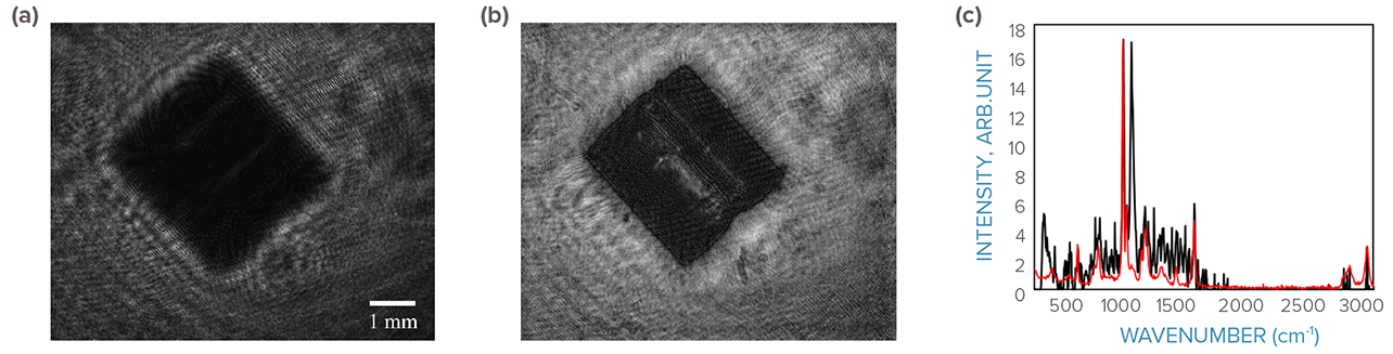
Figure 2. (a) Raw and (b) reconstructed images of a PS pellet. (c) Raman spectrum of a PS pellet taken using the setup (black) and reference sample in air (red). The sample peak at 1067 cm-¹ was attributed to AI2O3. Reprinted with permission from [1] © The Optical Society.
The creators believe that the hybrid system has the capacity to continuously monitor by utilizing holographic imaging to distinguish and locate particles, interrupting the flow at the appropriate time for Raman analysis when a particle of interest is detected.
Automation of this type would facilitate in-situ oceanographic monitoring of microplastics to better understand their distribution over time, and at a range of ocean depths – even those where particles are distributed sparsely.
Compared to the specifications of present-day holographic imaging systems for oceanographic particle detection, the innovative hybrid system offers an excellent balance between particle detection speed, flow throughput, and power consumption.
A Clever Match
Evaluating, understanding, and addressing the issues that microplastic pollution causes in our oceans demands international collaboration and continued innovation, as demonstrated in the development of this hybrid system for in-situ monitoring.
Digital holography could facilitate the automatic separation of microplastics from organic particles, while Raman spectroscopy delivers the specificity required for material identification.
Combining both methods with a single laser source and a compact, sensitive Raman spectrometer, the hybrid system can achieve size, speed, and power consumption requirements needed to make this a practical solution for in-situ monitoring of ocean microplastics.
References
- Takahashi, Tomoko, et al. (2020) “Identification of microplastics in a large water volume by integrated holography and Raman spectroscopy.” Applied Optics. 59,17, pp. 5073-5078.

This information has been sourced, reviewed and adapted from materials provided by Wasatch Photonics, Inc.
For more information on this source, please visit Wasatch Photonics, Inc.