Understanding the intricate structures and compositions of geological samples in Earth Science is central to research. This is particularly relevant in fields like carbon capture, utilization, and storage (CCUS), which begins with the capture of CO2 emissions, typically from power plants or industrial facilities. The captured gas, if not utilized on-site, is then compressed and transported for various applications or injected deep within geological formations, such as depleted oil reservoirs or saline aquifers.1
As our knowledge of geological formations grows and we develop more advanced energy solutions, tools capable of providing high-resolution insights are becoming increasingly valuable. At the forefront of scientific innovation, the Thermo Scientific Quattro ESEM Field Emission Environmental Scanning Electron Microscope (ESEM) allows for in-depth and versatile analysis of various geological materials, both in their natural state and under dynamic environmental conditions.
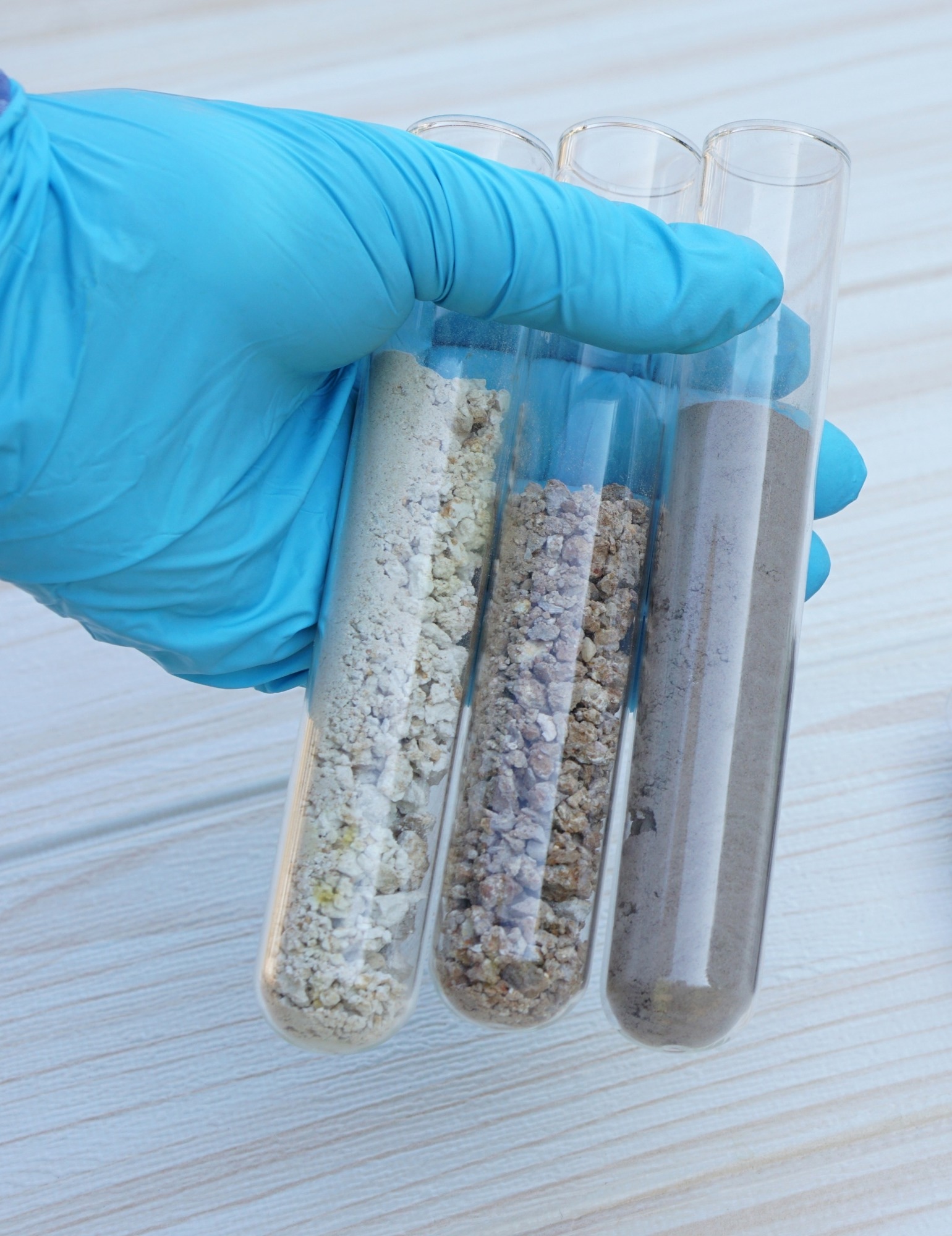
Image Credit: tawanroong/Shutterstock.com
Problem: When Traditional SEM Falls Short
Traditional scanning electron microscopes (SEMs) offer high-resolution imaging but require that samples be dehydrated before inspection. On particularly delicate samples, the dehydration process can cause structural damage owing to the force exerted by surface tension as the water evaporates, resulting in drying artifacts that alter the sample and hinder analysis. This limitation, in addition to requirements that the sample be static and conductive, means that essential characteristics, such as the wetting properties or dynamic changes of materials, cannot be adequately observed.2,3
In CCUS research, understanding how CO2, water, and rock interact in geological formations is critical. These relationships influence factors like water quality, mineral transformations, and the permeability of rocks, impacting the efficiency of CO2 sequestration. In failing to adequately capture dynamic and real-world conditions, conventional SEM limits the scope and scale of potential research.4
Challenges: Imaging Complex Geological Samples
Geological samples present unique challenges for electron-based imaging and analysis. Depending on the type of material, they can be porous, non-conductive, contain fluids, and react dynamically to environmental changes. Understanding these features, especially at the nanoscale, is vital for determining how rocks react to CO2 injections and for observing changes in wettability, pore structures, and mineral compositions.
Characterizing these complex samples also often requires transitioning between various imaging modes, which can be difficult especially under widely varying dynamic ranges. Thus, a multifaceted challenge lies in effectively imaging these samples while preserving their natural state.1
Solution: Unearthing the Full Picture with the Quattro ESEM
The Thermo Scientific Quattro ESEM offers a comprehensive solution with its impressive versatility and advanced imaging capabilities. Here is how it addresses the particular needs of Earth Science and CCUS research:
1. Environmental Scanning Electron Microscopy (ESEM) Capabilities
The Quattro ESEM is equipped with an advanced field emission gun (FEG) and unique environmental mode (ESEM) technology, allowing imaging in real-world conditions with minimal sample preparation. This grants researchers the ability to investigate hydrated, outgassing, and conductive/non-conductive samples while capturing dynamic changes such as wetting behavior.3
2. In Situ Analysis of Dynamic Changes
One of the standout features of the Quattro ESEM is its ability to perform in situ observations. The system can simulate various environmental conditions using water vapor and a temperature control stage, making it ideal for capturing how geological samples react to CO2, water, and other fluids in real-time. This is especially beneficial in CCUS research, where understanding mineral dissolution and precipitation, as well as changes in pore structures, is all-important.3
3. High-Resolution Imaging Across Various Scales
The Quattro ESEM supports high-resolution imaging from the micrometer to the nanometer scale. Its field emission electron source ensures exceptional resolution and image quality, while the system’s flexible, eucentric, 5-axis stage can accommodate a wide range of samples, from small rock chips to larger core samples. This flexibility allows for detailed analysis of rock structures, mineral compositions, and pore networks at various scales.3,4
4. Advanced Automation and Data Analysis with Maps Min and Avizo Software
The integration of Maps Min automated mineral analysis software and Avizo Software provides unparalleled imaging and data analysis capabilities.
Featuring a library consisting of thousands of mineral species, Maps Min Software delivers automated mineral identification, fast-tracking the analysis of complex samples. This was demonstrated in the lab when studying carbonate rock samples, where stitched, high-resolution images were produced to reveal intricate pore networks and mineral distributions.4,5
From the convenience of a single platform, Avizo Software provides optimized workflows for the characterization of advanced materials as well as quality control. It supports 2D/3D visualization and analysis, allowing researchers to correlate and quantify mineral changes across images.4,7
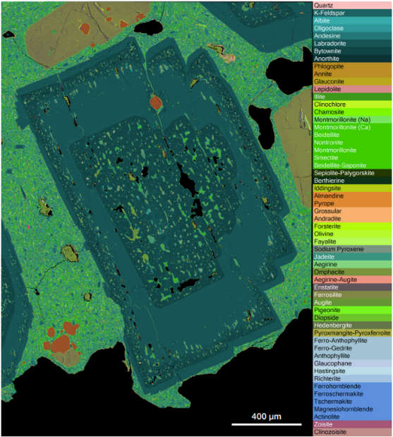
Maps Min analysis of geological sample. Image Credit: Thermo Fisher Scientific
5. Versatility in Vacuum Modes and Detectors
The Quattro ESEM features high-vacuum, low-vacuum, and ESEM modes, providing unmatched flexibility in accommodating a wide variety of samples. Its range of detectors in a high vacuum, including Everhart-Thornley, backscatter, and STEM detectors, supports comprehensive imaging and analytical capabilities. These detectors can direct the simultaneous acquisition of multiple signals, reducing beam exposure and enhancing data acquisition speed, which is particularly valuable when examining beam-sensitive or hydrated samples.3
Conclusion: Breaking New Ground in Earth Science Research
The Thermo Scientific Quattro ESEM represents a game-changing tool for Earth Science and CCUS research. Its ability to capture dynamic changes, perform high-resolution imaging, and provide comprehensive mineral analysis makes it a formidable instrument for researchers studying geological formations and material interactions under real-world conditions. By enabling the visualization of intricate pore structures, mineral compositions, and fluid dynamics, the Quattro ESEM guarantees that researchers gain a deeper understanding of geological materials, paving the way for advancements in CCUS technologies and beyond.
References and Further Reading
- Budinis, S., Fajardy, M. and Greenfield, C. (2024). Carbon Capture, Utilization and Storage - Energy System. (online) International Energy Agency. Available at: https://www.iea.org/energy-system/carbon-capture-utilisation-and-storage.
- Mudalige, T., et al. (2019). Characterization of Nanomaterials. Nanomaterials for Food Applications, pp.313–353. https://doi.org/10.1016/b978-0-12-814130-4.00011-7.
- ThermoFisher Scientific. Quattro ESEM Ultra-versatile, high-resolution SEM with unique environmental capability. Available at: https://assets.thermofisher.com/TFS-Assets/MSD/brochures/quattro-brochure-br0148-en.pdf (Accessed 20 May 2025).
- Thermo Scientific. (2024). Quattro ESEM CCUS Application Brief.
- ThermoFisher Scientific.(2025). Mining Industry | Mineralogy Software | Thermo Fisher Scientific - US. (online) Available at: https://www.thermofisher.com/ca/en/home/electron-microscopy/products/software-em-3d-vis/maps-min.html (Accessed 20 May 2025).
- Hanna, R.D. and Ketcham, R.A. (2017). X-ray computed tomography of planetary materials: A primer and review of recent studies. 77(4), pp.547–572. https://doi.org/10.1016/j.chemer.2017.01.006.
- ThermoFisher Scientific. (2025). Avizo Software | Materials Characterization Software | Thermo Fisher Scientific - US. (online) Available at: https://www.thermofisher.com/ca/en/home/electron-microscopy/products/software-em-3d-vis/avizo-software.html (Accessed 20 May 2025).

This information has been sourced, reviewed and adapted from materials provided by Thermo Fisher Scientific – Electron Microscopy Solutions North America.
For more information on this source, please visit Thermo Fisher Scientific – Electron Microscopy Solutions North America.